 Movie
Movie Controller

[English] 日本語
 Yorodumi
- PDB-5c7o: Structure of human testis-specific glyceraldehyde-3-phosphate deh... -
+ Open data

- Basic information
| Entry | Database: PDB / ID: 5c7o | ||||||
|---|---|---|---|---|---|---|---|
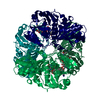
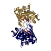
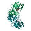
| Title | Structure of human testis-specific glyceraldehyde-3-phosphate dehydrogenase holo form with NAD+ | ||||||
 Components Components | Glyceraldehyde-3-phosphate dehydrogenase, testis-specific Glyceraldehyde 3-phosphate dehydrogenase Glyceraldehyde 3-phosphate dehydrogenase | ||||||
 Keywords Keywords | OXIDOREDUCTASE / human sperm GAPDH metabolic enzyme sperm specific with NAD+ cofactor | ||||||
| Function / homology |  Function and homology information Function and homology informationflagellated sperm motility / glyceraldehyde-3-phosphate dehydrogenase (phosphorylating) / glyceraldehyde-3-phosphate dehydrogenase (NAD+) (phosphorylating) activity / Gluconeogenesis / Glycolysis / Association of TriC/CCT with target proteins during biosynthesis / positive regulation of glycolytic process / glycolytic process / glucose metabolic process / NAD binding ...flagellated sperm motility / glyceraldehyde-3-phosphate dehydrogenase (phosphorylating) / glyceraldehyde-3-phosphate dehydrogenase (NAD+) (phosphorylating) activity / Gluconeogenesis / Glycolysis / Association of TriC/CCT with target proteins during biosynthesis / positive regulation of glycolytic process / glycolytic process / glucose metabolic process / NAD binding / NADP binding / nucleus / cytosolSimilarity search - Function | ||||||
| Biological species |  Homo sapiens (human) Homo sapiens (human) | ||||||
| Method | X-RAY DIFFRACTION / SYNCHROTRON / MOLECULAR REPLACEMENT / Resolution: 1.729 Å | ||||||
 Authors Authors | Betts, L. / Machius, M. / Danshina, P. / O'Brien, D. | ||||||
 Citation Citation | Journal: Mol. Hum. Reprod. / Year: 2016 Title: Structural analyses to identify selective inhibitors of glyceraldehyde 3-phosphate dehydrogenase-S, a sperm-specific glycolytic enzyme. Authors: Danshina, P.V. / Qu, W. / Temple, B.R. / Rojas, R.J. / Miley, M.J. / Machius, M. / Betts, L. / O'Brien, D.A. | ||||||
| History |
|
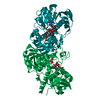
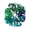
- Structure visualization
| Structure viewer | Molecule: MolmilJmol/JSmol |
|---|
- Downloads & links
-Download
| PDBx/mmCIF format | 5c7o.cif.gz | 270.6 KB | Display | PDBx/mmCIF format |
|---|---|---|---|---|
| PDB format | pdb5c7o.ent.gz | 219.9 KB | Display | PDB format |
| PDBx/mmJSON format | 5c7o.json.gz | Tree view | PDBx/mmJSON format | |
| Others |  Other downloads Other downloads |
-Validation report
| Arichive directory | https://data.pdbj.org/pub/pdb/validation_reports/c7/5c7oftp://data.pdbj.org/pub/pdb/validation_reports/c7/5c7o | HTTPS FTP |
|---|
-Related structure data
| Related structure data |  5c7iC 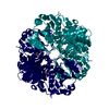 5c7lC 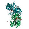 3h9eS C: citing same article ( S: Starting model for refinement |
|---|---|
| Similar structure data |


-Links
 PDBj
PDBj


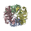
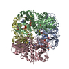
- Assembly
| Deposited unit | 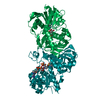
| ||||||||||||||||||
|---|---|---|---|---|---|---|---|---|---|---|---|---|---|---|---|---|---|---|---|
| 1 | 
| ||||||||||||||||||
| Unit cell |
| ||||||||||||||||||
| Components on special symmetry positions |
|

-Components
| #1: Protein | Glyceraldehyde 3-phosphate dehydrogenase / Spermatogenic cell-specific glyceraldehyde 3-phosphate dehydrogenase 2 / GAPDH-2 / Spermatogenic ...Spermatogenic cell-specific glyceraldehyde 3-phosphate dehydrogenase 2 / GAPDH-2 / Spermatogenic glyceraldehyde-3-phosphate dehydrogenase Mass: 36667.746 Da / Num. of mol.: 2 / Fragment: unp residues 74-407 Source method: isolated from a genetically manipulated source Source: (gene. exp.) Homo sapiens (human) / Gene: GAPDHS, GAPD2, GAPDH2, GAPDS, HSD-35, HSD35 / Production host:  Escherichia coli (E. coli) Escherichia coli (E. coli)References: UniProt: O14556, glyceraldehyde-3-phosphate dehydrogenase (phosphorylating)#2: Chemical | Nicotinamide adenine dinucleotide  Mass: 663.425 Da / Num. of mol.: 2 / Source method: obtained synthetically / Formula: C21H27N7O14P2 / Comment: NAD*YM Mass: 663.425 Da / Num. of mol.: 2 / Source method: obtained synthetically / Formula: C21H27N7O14P2 / Comment: NAD*YM#3: Chemical | Sulfate  Mass: 96.063 Da / Num. of mol.: 2 / Source method: obtained synthetically / Formula: SO4 Mass: 96.063 Da / Num. of mol.: 2 / Source method: obtained synthetically / Formula: SO4#4: Water | ChemComp-HOH / | Water Mass: 18.015 Da / Num. of mol.: 629 / Source method: isolated from a natural source / Formula: H2O Mass: 18.015 Da / Num. of mol.: 629 / Source method: isolated from a natural source / Formula: H2O |
|---|
-Experimental details
-Experiment
| Experiment | Method: X-RAY DIFFRACTION / Number of used crystals: 1 |
|---|
- Sample preparation
| Crystal | Density Matthews: 2.4 Å3/Da / Density % sol: 48.74 % |
|---|---|
| Crystal grow | Temperature: 273 K / Method: vapor diffusion Details: 1:1 mixture of protein:NAD+ complex at 10 mg/mL with 0.2 M sodium nitrate, 20% PEG3350 |
-Data collection
| Diffraction | Mean temperature: 100 K |
|---|---|
| Diffraction source | Source: SYNCHROTRON / Site: APS  / Beamline: 22-ID / Wavelength: 1.0746 Å / Beamline: 22-ID / Wavelength: 1.0746 Å |
| Detector | Type: MARMOSAIC 300 mm CCD / Detector: CCD / Date: Apr 10, 2014 |
| Radiation | Protocol: SINGLE WAVELENGTH / Monochromatic (M) / Laue (L): M / Scattering type: x-ray |
| Radiation wavelength | Wavelength: 1.0746 Å / Relative weight: 1 |
| Reflection | Resolution: 1.73→31.71 Å / Num. obs: 68115 / % possible obs: 93.9 % / Redundancy: 4.2 % / Net I/σ(I): 19.2 |
- Processing
| Software |
| |||||||||||||||||||||||||||||||||||||||||||||||||||||||||||||||||||||||||||||||||||||||||||||||||||||||||
|---|---|---|---|---|---|---|---|---|---|---|---|---|---|---|---|---|---|---|---|---|---|---|---|---|---|---|---|---|---|---|---|---|---|---|---|---|---|---|---|---|---|---|---|---|---|---|---|---|---|---|---|---|---|---|---|---|---|---|---|---|---|---|---|---|---|---|---|---|---|---|---|---|---|---|---|---|---|---|---|---|---|---|---|---|---|---|---|---|---|---|---|---|---|---|---|---|---|---|---|---|---|---|---|---|---|---|
| Refinement | Method to determine structure: MOLECULAR REPLACEMENT Starting model: 3H9E Resolution: 1.729→31.706 Å / SU ML: 0.15 / Cross valid method: FREE R-VALUE / σ(F): 1.34 / Phase error: 16.51 / Stereochemistry target values: ML
| |||||||||||||||||||||||||||||||||||||||||||||||||||||||||||||||||||||||||||||||||||||||||||||||||||||||||
| Solvent computation | Shrinkage radii: 0.9 Å / VDW probe radii: 1.11 Å / Solvent model: FLAT BULK SOLVENT MODEL | |||||||||||||||||||||||||||||||||||||||||||||||||||||||||||||||||||||||||||||||||||||||||||||||||||||||||
| Displacement parameters | Biso max: 96.93 Å2 / Biso mean: 26.8695 Å2 / Biso min: 10.59 Å2 | |||||||||||||||||||||||||||||||||||||||||||||||||||||||||||||||||||||||||||||||||||||||||||||||||||||||||
| Refinement step | Cycle: final / Resolution: 1.729→31.706 Å
| |||||||||||||||||||||||||||||||||||||||||||||||||||||||||||||||||||||||||||||||||||||||||||||||||||||||||
| Refine LS restraints |
| |||||||||||||||||||||||||||||||||||||||||||||||||||||||||||||||||||||||||||||||||||||||||||||||||||||||||
| LS refinement shell | Refine-ID: X-RAY DIFFRACTION / Total num. of bins used: 14
|
