 Movie
Movie Controller


+ Open data

- Basic information
| Entry | Database: PDB / ID: 6wjh | ||||||
|---|---|---|---|---|---|---|---|
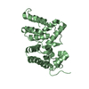
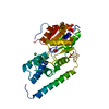
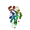
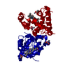
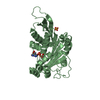
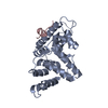
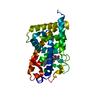
| Title | Crystal structure of MAGE-A11 bound to the PCF11 degron | ||||||
 Components Components | Fusion protein of PCF11 and MAGE-A11 | ||||||
 Keywords Keywords | LIGASE / MAGE-A11 / PCF 11 / ubiquitin ligase / protein complex | ||||||
| Function / homology |  Function and homology information: / mRNA cleavage factor complex / Processing of Intronless Pre-mRNAs / mRNA 3'-end processing / mRNA 3'-end processing / RNA Polymerase II Transcription Termination / : / termination of RNA polymerase II transcription / RNA polymerase II complex binding / RNA polymerase II transcribes snRNA genes ...: / mRNA cleavage factor complex / Processing of Intronless Pre-mRNAs / mRNA 3'-end processing / mRNA 3'-end processing / RNA Polymerase II Transcription Termination / : / termination of RNA polymerase II transcription / RNA polymerase II complex binding / RNA polymerase II transcribes snRNA genes / mRNA Splicing - Major Pathway / mRNA splicing, via spliceosome / nuclear body / mRNA binding / mitochondrion / nucleoplasm / cytosol / cytoplasm Function and homology information: / mRNA cleavage factor complex / Processing of Intronless Pre-mRNAs / mRNA 3'-end processing / mRNA 3'-end processing / RNA Polymerase II Transcription Termination / : / termination of RNA polymerase II transcription / RNA polymerase II complex binding / RNA polymerase II transcribes snRNA genes ...: / mRNA cleavage factor complex / Processing of Intronless Pre-mRNAs / mRNA 3'-end processing / mRNA 3'-end processing / RNA Polymerase II Transcription Termination / : / termination of RNA polymerase II transcription / RNA polymerase II complex binding / RNA polymerase II transcribes snRNA genes / mRNA Splicing - Major Pathway / mRNA splicing, via spliceosome / nuclear body / mRNA binding / mitochondrion / nucleoplasm / cytosol / cytoplasmSimilarity search - Function | ||||||
| Biological species |  Homo sapiens (human) Homo sapiens (human) | ||||||
| Method | X-RAY DIFFRACTION / SYNCHROTRON / MOLECULAR REPLACEMENT / molecular replacement / Resolution: 2.19 Å | ||||||
 Authors Authors | Miller, D.J. / Huang, X. | ||||||
| Funding support |  United States, 1items United States, 1items
| ||||||
 Citation Citation | Journal: Nat Commun / Year: 2020 Title: Structural basis for substrate recognition and chemical inhibition of oncogenic MAGE ubiquitin ligases. Authors: Yang, S.W. / Huang, X. / Lin, W. / Min, J. / Miller, D.J. / Mayasundari, A. / Rodrigues, P. / Griffith, E.C. / Gee, C.T. / Li, L. / Li, W. / Lee, R.E. / Rankovic, Z. / Chen, T. / Potts, P.R. | ||||||
| History |
|
- Structure visualization
| Structure viewer | Molecule: MolmilJmol/JSmol |
|---|
- Downloads & links
-Download
| PDBx/mmCIF format | 6wjh.cif.gz | 189.7 KB | Display | PDBx/mmCIF format |
|---|---|---|---|---|
| PDB format | pdb6wjh.ent.gz | 149.5 KB | Display | PDB format |
| PDBx/mmJSON format | 6wjh.json.gz | Tree view | PDBx/mmJSON format | |
| Others |  Other downloads Other downloads |
-Validation report
| Arichive directory | https://data.pdbj.org/pub/pdb/validation_reports/wj/6wjhftp://data.pdbj.org/pub/pdb/validation_reports/wj/6wjh | HTTPS FTP |
|---|
-Related structure data
| Related structure data | 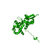 4v0pS S: Starting model for refinement |
|---|---|
| Similar structure data |


-Links
 PDBj
PDBj


- Assembly
| Deposited unit | 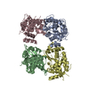
| ||||||||
|---|---|---|---|---|---|---|---|---|---|
| 1 | 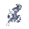
| ||||||||
| 2 | 
| ||||||||
| 3 | 
| ||||||||
| 4 | 
| ||||||||
| Unit cell |
|
-Components
| #1: Protein | / Pre-mRNA cleavage complex II protein Pcf11 / Cancer/testis antigen 1.11 / CT1.11 / MAGE-11 antigen Mass: 27800.822 Da / Num. of mol.: 4 Source method: isolated from a genetically manipulated source Source: (gene. exp.) Homo sapiens (human) / Gene: PCF11, KIAA0824, MAGEA11, MAGE11 / Production host:  Escherichia coli (E. coli) / References: UniProt: O94913, UniProt: P43364 Escherichia coli (E. coli) / References: UniProt: O94913, UniProt: P43364#2: Water | ChemComp-HOH / | Water Mass: 18.015 Da / Num. of mol.: 208 / Source method: isolated from a natural source / Formula: H2O Mass: 18.015 Da / Num. of mol.: 208 / Source method: isolated from a natural source / Formula: H2O |
|---|
-Experimental details
-Experiment
| Experiment | Method: X-RAY DIFFRACTION / Number of used crystals: 1 |
|---|
- Sample preparation
| Crystal | Density Matthews: 2.71 Å3/Da / Density % sol: 54.56 % |
|---|---|
| Crystal grow | Temperature: 293.15 K / Method: vapor diffusion, sitting drop Details: The well solution contained 0.2 M Na acetate, 0.1 M Bis Tris propane pH 7.5 and 20% (w/v) PEG 3350. The drop contained a 1:1 volume ratio of well solution to protein (15 mg/ml in 50 mM Tris, ...Details: The well solution contained 0.2 M Na acetate, 0.1 M Bis Tris propane pH 7.5 and 20% (w/v) PEG 3350. The drop contained a 1:1 volume ratio of well solution to protein (15 mg/ml in 50 mM Tris, pH 7.4, 200 mM NaCl and 5 mM DTT). |
-Data collection
| Diffraction | Mean temperature: 100 K / Serial crystal experiment: N |
|---|---|
| Diffraction source | Source: SYNCHROTRON / Site: APS / Beamline: 22-BM / Wavelength: 1 Å |
| Detector | Type: RAYONIX MX300-HS / Detector: CCD / Date: Nov 1, 2018 |
| Radiation | Protocol: SINGLE WAVELENGTH / Monochromatic (M) / Laue (L): M / Scattering type: x-ray |
| Radiation wavelength | Wavelength: 1 Å / Relative weight: 1 |
| Reflection | Resolution: 2.19→38.79 Å / Num. obs: 56569 / % possible obs: 96.6 % / Observed criterion σ(I): -3 / Redundancy: 2.4 % / CC1/2: 0.952 / Rsym value: 0.12 / Net I/σ(I): 19.9 |
| Reflection shell | Resolution: 2.19→2.25 Å / Num. unique obs: 2745 / CC1/2: 0.757 / Rsym value: 0.372 |
-Phasing
| Phasing | Method: molecular replacement |
|---|
- Processing
| Software |
| ||||||||||||||||||||||||||||||||||||||||||||||||||||||||||||
|---|---|---|---|---|---|---|---|---|---|---|---|---|---|---|---|---|---|---|---|---|---|---|---|---|---|---|---|---|---|---|---|---|---|---|---|---|---|---|---|---|---|---|---|---|---|---|---|---|---|---|---|---|---|---|---|---|---|---|---|---|---|
| Refinement | Method to determine structure: MOLECULAR REPLACEMENT Starting model: 4V0P Resolution: 2.19→38.79 Å / Cor.coef. Fo:Fc: 0.945 / Cor.coef. Fo:Fc free: 0.925 / SU B: 6.162 / SU ML: 0.157 / Cross valid method: THROUGHOUT / σ(F): 0 / ESU R: 0.254 / ESU R Free: 0.206 Details: HYDROGENS HAVE BEEN ADDED IN THE RIDING POSITIONS U VALUES : REFINED INDIVIDUALLY
| ||||||||||||||||||||||||||||||||||||||||||||||||||||||||||||
| Solvent computation | Ion probe radii: 0.8 Å / Shrinkage radii: 0.8 Å / VDW probe radii: 1.2 Å | ||||||||||||||||||||||||||||||||||||||||||||||||||||||||||||
| Displacement parameters | Biso max: 94.55 Å2 / Biso mean: 36.814 Å2 / Biso min: 21.59 Å2
| ||||||||||||||||||||||||||||||||||||||||||||||||||||||||||||
| Refinement step | Cycle: final / Resolution: 2.19→38.79 Å
| ||||||||||||||||||||||||||||||||||||||||||||||||||||||||||||
| Refine LS restraints |
| ||||||||||||||||||||||||||||||||||||||||||||||||||||||||||||
| LS refinement shell | Resolution: 2.194→2.251 Å / Rfactor Rfree error: 0 / Total num. of bins used: 20
|
