 Movie
Movie Controller

[English] 日本語
 Yorodumi
- PDB-6wit: Crystal structure of NHP D15.SD7 Fab in complex with 16055 V1V2 1... -
+ Open data

- Basic information
| Entry | Database: PDB / ID: 6wit | ||||||
|---|---|---|---|---|---|---|---|
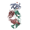
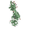
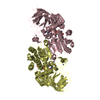
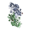
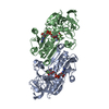
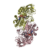
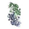
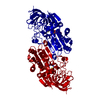
| Title | Crystal structure of NHP D15.SD7 Fab in complex with 16055 V1V2 1FD6 scaffold | ||||||
 Components Components |
| ||||||
 Keywords Keywords |  ANTIVIRAL PROTEIN/IMMUNE SYSTEM / Fab / HIV / V1V2 / Complex / Antibody / Scaffold / ANTIVIRAL PROTEIN-IMMUNE SYSTEM complex ANTIVIRAL PROTEIN/IMMUNE SYSTEM / Fab / HIV / V1V2 / Complex / Antibody / Scaffold / ANTIVIRAL PROTEIN-IMMUNE SYSTEM complex | ||||||
| Biological species |  Macaca mulatta (Rhesus monkey) Macaca mulatta (Rhesus monkey) Homo sapiens (human) Homo sapiens (human) | ||||||
| Method | X-RAY DIFFRACTION / SYNCHROTRON / MOLECULAR REPLACEMENT / Resolution: 2.79 Å | ||||||
 Authors Authors | Liban, T. / Aljedani, S. / Rodarte, J. / Pancera, M. | ||||||
| Funding support |  United States, 1items United States, 1items
| ||||||
 Citation Citation | Journal: Plos Pathog. / Year: 2021 Title: Structurally related but genetically unrelated antibody lineages converge on an immunodominant HIV-1 Env neutralizing determinant following trimer immunization. Authors: Aljedani, S.S. / Liban, T.J. / Tran, K. / Phad, G. / Singh, S. / Dubrovskaya, V. / Pushparaj, P. / Martinez-Murillo, P. / Rodarte, J. / Mileant, A. / Mangala Prasad, V. / Kinzelman, R. / ...Authors: Aljedani, S.S. / Liban, T.J. / Tran, K. / Phad, G. / Singh, S. / Dubrovskaya, V. / Pushparaj, P. / Martinez-Murillo, P. / Rodarte, J. / Mileant, A. / Mangala Prasad, V. / Kinzelman, R. / O'Dell, S. / Mascola, J.R. / Lee, K.K. / Karlsson Hedestam, G.B. / Wyatt, R.T. / Pancera, M. | ||||||
| History |
|
- Structure visualization
| Structure viewer | Molecule:  MolmilJmol/JSmol MolmilJmol/JSmol |
|---|
- Downloads & links
-Download
| PDBx/mmCIF format | 6wit.cif.gz | 388.1 KB | Display | PDBx/mmCIF format |
|---|---|---|---|---|
| PDB format | pdb6wit.ent.gz | 318.9 KB | Display | PDB format |
| PDBx/mmJSON format | 6wit.json.gz | Tree view | PDBx/mmJSON format | |
| Others |  Other downloads Other downloads |
-Validation report
| Arichive directory | https://data.pdbj.org/pub/pdb/validation_reports/wi/6witftp://data.pdbj.org/pub/pdb/validation_reports/wi/6wit | HTTPS FTP |
|---|
-Related structure data
| Related structure data |  6vjnC  6wasC  6xlzC  6xsnC  4rfoS C: citing same article ( S: Starting model for refinement |
|---|---|
| Similar structure data |
-Links
 PDBj
PDBj


- Assembly
| Deposited unit | 
| ||||||||
|---|---|---|---|---|---|---|---|---|---|
| 1 | 
| ||||||||
| 2 | 
| ||||||||
| Unit cell |
|
-Components
-Antibody , 2 types, 4 molecules AHBL
| #1: Antibody | Mass: 25107.119 Da / Num. of mol.: 2 Source method: isolated from a genetically manipulated source Source: (gene. exp.) Macaca mulatta (Rhesus monkey) / Cell (production host): 293e / Production host: Homo sapiens (human)#2: Antibody | Mass: 22793.998 Da / Num. of mol.: 2 Source method: isolated from a genetically manipulated source Source: (gene. exp.) Macaca mulatta (Rhesus monkey) / Cell (production host): 293e / Production host: Homo sapiens (human) |
|---|
-Protein / Sugars , 2 types, 6 molecules CI
| #3: Protein | Mass: 13995.898 Da / Num. of mol.: 2 Source method: isolated from a genetically manipulated source Source: (gene. exp.) Homo sapiens (human) / Cell (production host): 293e / Production host: Homo sapiens (human)#4: Sugar | ChemComp-NAG / N-Acetylglucosamine Type: D-saccharide, beta linking / Mass: 221.208 Da / Num. of mol.: 4 Type: D-saccharide, beta linking / Mass: 221.208 Da / Num. of mol.: 4Source method: isolated from a genetically manipulated source Formula: C8H15NO6 |
|---|
-Non-polymers , 2 types, 75 molecules 

| #5: Chemical | ChemComp-SO4 / Sulfate Mass: 96.063 Da / Num. of mol.: 4 / Source method: obtained synthetically / Formula: SO4 Mass: 96.063 Da / Num. of mol.: 4 / Source method: obtained synthetically / Formula: SO4#6: Water | ChemComp-HOH / | WaterMass: 18.015 Da / Num. of mol.: 71 / Source method: isolated from a natural source / Formula: H2O |
|---|
-Details
| Has ligand of interest | N |
|---|
-Experimental details
-Experiment
| Experiment | Method: X-RAY DIFFRACTION / Number of used crystals: 1 |
|---|
- Sample preparation
| Crystal | Density Matthews: 2.33 Å3/Da / Density % sol: 47.25 % |
|---|---|
| Crystal grow | Temperature: 293.15 K / Method: vapor diffusion, hanging drop Details: 0.2 M ammonium sulfate, 0.1 M MES, pH 6.5, 22% PEG8000 |
-Data collection
| Diffraction | Mean temperature: 100 K / Serial crystal experiment: N |
|---|---|
| Diffraction source | Source: SYNCHROTRON / Site: ALS / Beamline: 5.0.1 / Wavelength: 1 Å |
| Detector | Type: DECTRIS PILATUS3 6M / Detector: PIXEL / Date: Dec 21, 2017 |
| Radiation | Monochromator: double crystal Si(111) / Protocol: SINGLE WAVELENGTH / Monochromatic (M) / Laue (L): M / Scattering type: x-ray |
| Radiation wavelength | Wavelength: 1 Å / Relative weight: 1 |
| Reflection | Resolution: 2.79→48.4813 Å / Num. obs: 28183 / % possible obs: 95.72 % / Redundancy: 6.2 % / Biso Wilson estimate: 49.64 Å2 / CC1/2: 0.992 / CC star: 0.998 / Rmerge(I) obs: 0.144 / Rpim(I) all: 0.049 / Rrim(I) all: 0.125 / Rsym value: 0.084 / Χ2: 0.936 / Net I/σ(I): 16.05 |
| Reflection shell | Resolution: 2.8→2.85 Å / Redundancy: 4.2 % / Rmerge(I) obs: 0.927 / Mean I/σ(I) obs: 1.47 / Num. unique obs: 1436 / CC1/2: 0.573 / CC star: 0.853 / Rpim(I) all: 0.573 / Rrim(I) all: 0.495 / Rsym value: 0.408 / % possible all: 66.75 |
- Processing
| Software |
| |||||||||||||||||||||||||||||||||||||||||||||||||||||||||||||||||||||||||||||||||||||||||||||||||||||||||
|---|---|---|---|---|---|---|---|---|---|---|---|---|---|---|---|---|---|---|---|---|---|---|---|---|---|---|---|---|---|---|---|---|---|---|---|---|---|---|---|---|---|---|---|---|---|---|---|---|---|---|---|---|---|---|---|---|---|---|---|---|---|---|---|---|---|---|---|---|---|---|---|---|---|---|---|---|---|---|---|---|---|---|---|---|---|---|---|---|---|---|---|---|---|---|---|---|---|---|---|---|---|---|---|---|---|---|
| Refinement | Method to determine structure: MOLECULAR REPLACEMENT Starting model: PDB entry 4RFO Resolution: 2.79→48.48 Å / SU ML: 0.32 / Cross valid method: FREE R-VALUE / σ(F): 1.34 / Phase error: 29.06 / Stereochemistry target values: ML
| |||||||||||||||||||||||||||||||||||||||||||||||||||||||||||||||||||||||||||||||||||||||||||||||||||||||||
| Solvent computation | Shrinkage radii: 0.9 Å / VDW probe radii: 1.11 Å / Solvent model: FLAT BULK SOLVENT MODEL | |||||||||||||||||||||||||||||||||||||||||||||||||||||||||||||||||||||||||||||||||||||||||||||||||||||||||
| Refinement step | Cycle: LAST / Resolution: 2.79→48.48 Å
| |||||||||||||||||||||||||||||||||||||||||||||||||||||||||||||||||||||||||||||||||||||||||||||||||||||||||
| Refine LS restraints |
| |||||||||||||||||||||||||||||||||||||||||||||||||||||||||||||||||||||||||||||||||||||||||||||||||||||||||
| LS refinement shell |
|
