[English] 日本語
Yorodumi
- PDB-6uug: Structure of methanesulfinate monooxygenase MsuC from Pseudomonas... -

+
Open data


ID or keywords:

Loading...

-
Basic information

Entry
Database: PDB / ID: 6uug
TitleStructure of methanesulfinate monooxygenase MsuC from Pseudomonas fluorescens at 1.69 angstrom resolution
ComponentsPutative dehydrogenase
KeywordsFLAVOPROTEIN / two-component flavin-dependent monooxygenase / methyl sulfur assimilation / dimethylsulfide / global sulfur cycle
Function / homology
Function and homology information


oxidoreductase activity, acting on the CH-CH group of donors / monooxygenase activity / flavin adenine dinucleotide binding
Similarity search - Function
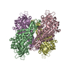
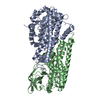
Acyl-CoA dehydrogenase, C-terminal domain / Acyl-CoA dehydrogenase, C-terminal domain / Butyryl-Coa Dehydrogenase, subunit A; domain 1 / Acyl-CoA dehydrogenase/oxidase, N-terminal domain / Acyl-CoA oxidase/dehydrogenase, middle domain superfamily / Butyryl-CoA Dehydrogenase, subunit A, domain 2 / Butyryl-CoA Dehydrogenase, subunit A; domain 2 / Acyl-CoA dehydrogenase/oxidase, N-terminal / Acyl-CoA dehydrogenase, N-terminal domain / Acyl-CoA dehydrogenase/oxidase, N-terminal domain superfamily ...Acyl-CoA dehydrogenase, C-terminal domain / Acyl-CoA dehydrogenase, C-terminal domain / Butyryl-Coa Dehydrogenase, subunit A; domain 1 / Acyl-CoA dehydrogenase/oxidase, N-terminal domain / Acyl-CoA oxidase/dehydrogenase, middle domain superfamily / Butyryl-CoA Dehydrogenase, subunit A, domain 2 / Butyryl-CoA Dehydrogenase, subunit A; domain 2 / Acyl-CoA dehydrogenase/oxidase, N-terminal / Acyl-CoA dehydrogenase, N-terminal domain / Acyl-CoA dehydrogenase/oxidase, N-terminal domain superfamily / Butyryl-CoA Dehydrogenase, subunit A, domain 3 / Acyl-CoA dehydrogenase/oxidase, N-terminal and middle domain superfamily / Acyl-CoA dehydrogenase-like, C-terminal / Butyryl-CoA Dehydrogenase, subunit A; domain 3 / Up-down Bundle / Beta Barrel / Orthogonal Bundle / Mainly Beta / Mainly Alpha
Similarity search - Domain/homology
Putative dehydrogenase
Similarity search - Component
Biological speciesPseudomonas fluorescens (bacteria)
MethodX-RAY DIFFRACTION / SYNCHROTRON / MOLECULAR REPLACEMENT / Resolution: 1.685 Å
AuthorsSoule, J. / Gnann, A.D. / Gonzalez, R. / Parker, M.J. / McKenna, K.C. / Nguyen, S.V. / Phan, N.T. / Wicht, D.K. / Dowling, D.P.
Funding support United States, 1items
OrganizationGrant numberCountry
National Science Foundation (NSF, United States)1807480 United States
CitationJournal: Biochem.Biophys.Res.Commun. / Year: 2020
Title: Structure and function of the two-component flavin-dependent methanesulfinate monooxygenase within bacterial sulfur assimilation.
Authors: Soule, J. / Gnann, A.D. / Gonzalez, R. / Parker, M.J. / McKenna, K.C. / Nguyen, S.V. / Phan, N.T. / Wicht, D.K. / Dowling, D.P.
History
DepositionOct 30, 2019Deposition site: RCSB / Processing site: RCSB
Revision 1.0Dec 4, 2019Provider: repository / Type: Initial release
Revision 1.1Jan 8, 2020Group: Author supporting evidence / Category: pdbx_audit_support / Item: _pdbx_audit_support.funding_organization
Revision 1.2Jan 22, 2020Group: Database references / Category: citation
Item: _citation.journal_volume / _citation.page_first ..._citation.journal_volume / _citation.page_first / _citation.page_last / _citation.year
Revision 1.3May 6, 2020Group: Structure summary / Category: audit_author / Item: _audit_author.name
Revision 1.4Oct 11, 2023Group: Data collection / Database references / Refinement description
Category: chem_comp_atom / chem_comp_bond ...chem_comp_atom / chem_comp_bond / database_2 / pdbx_initial_refinement_model
Item: _database_2.pdbx_DOI / _database_2.pdbx_database_accession

-
Structure visualization

Structure viewerMolecule:
MolmilJmol/JSmol

Downloads & links

-
Assembly

Deposited unit
A: Putative dehydrogenase
B: Putative dehydrogenase


Theoretical massNumber of molelcules
Total (without water)92,5162
Polymers92,5162
Non-polymers00
Water6,684371
1
A: Putative dehydrogenase
B: Putative dehydrogenase

A: Putative dehydrogenase
B: Putative dehydrogenase


Theoretical massNumber of molelcules
Total (without water)185,0324
Polymers185,0324
Non-polymers00
Water724
TypeNameSymmetry operationNumber
identity operation1_555x,y,z1
crystal symmetry operation2_555-x,-y,z1
Buried area13600 Å2
ΔGint-95 kcal/mol
Surface area53320 Å2
MethodPISA
Unit cell
Length a, b, c (Å)81.187, 161.582, 62.045
Angle α, β, γ (deg.)90.000, 90.000, 90.000
Int Tables number18
Space group name H-MP21212
Space group name HallP22ab
Symmetry operation#1: x,y,z
#2: x+1/2,-y+1/2,-z
#3: -x+1/2,y+1/2,-z
#4: -x,-y,z

-
Components

#1: Protein Putative dehydrogenase /


Mass: 46258.012 Da / Num. of mol.: 2
Source method: isolated from a genetically manipulated source
Source: (gene. exp.) Pseudomonas fluorescens (strain Pf0-1) (bacteria)
Strain: Pf0-1 / Gene: Pfl01_3917 / Plasmid: pPfl01_msuC / Production host: Escherichia coli BL21(DE3) (bacteria) / References: UniProt: Q3K9A0
#2: Water ChemComp-HOH / water / Water


Mass: 18.015 Da / Num. of mol.: 371 / Source method: isolated from a natural source / Formula: H2O

-
Experimental details

-
Experiment

ExperimentMethod: X-RAY DIFFRACTION / Number of used crystals: 1

-
Sample preparation

CrystalDensity Matthews: 2.28 Å3/Da / Density % sol: 45.95 % / Description: trapezoidal shaped, small crystals
Crystal growTemperature: 291 K / Method: vapor diffusion, sitting drop / pH: 8.5
Details: 0.1 M Tris (pH 8.5) 0.15 M CaCl2 30% (w/v) PEG 3350

-
Data collection

DiffractionMean temperature: 100 K / Serial crystal experiment: N
Diffraction sourceSource: SYNCHROTRON / Site: APS / Beamline: 24-ID-E / Wavelength: 0.9792 Å
DetectorType: DECTRIS EIGER X 16M / Detector: PIXEL / Date: Aug 20, 2019
RadiationProtocol: SINGLE WAVELENGTH / Monochromatic (M) / Laue (L): M / Scattering type: x-ray
Radiation wavelengthWavelength: 0.9792 Å / Relative weight: 1
ReflectionResolution: 1.68→62.045 Å / Num. obs: 92686 / % possible obs: 99.7 % / Redundancy: 6.6 % / Biso Wilson estimate: 42.2 Å2 / CC1/2: 1 / Rpim(I) all: 0.017 / Rrim(I) all: 0.043 / Rsym value: 0.041 / Net I/σ(I): 20.32
Reflection shellResolution: 1.68→1.79 Å / Redundancy: 6.7 % / Mean I/σ(I) obs: 1.42 / Num. unique obs: 14648 / CC1/2: 0.655 / Rpim(I) all: 0.479 / Rsym value: 1.164 / % possible all: 98.7

-
Processing

Software
NameVersionClassification
PHENIX1.15.2_3472refinement
XDSVERSION Mar 15, 2019data reduction
XDSVERSION Mar 15, 2019data scaling
PHASER2.5.6phasing
Coot0.8.9.1model building
RefinementMethod to determine structure: MOLECULAR REPLACEMENT
Starting model: 5XB8
Resolution: 1.685→62.045 Å / Cross valid method: FREE R-VALUE
Details: Rounds of simulated annealing, minimization, and B-factor refinement were conducted until convergence. Occupancies of alternate side-chain conformations were optimized towards the end of refinement.
RfactorNum. reflection% reflection
Rfree0.2082 4633 5 %
Rwork0.1852 --
obs0.1864 87979 99.69 %
Solvent computationShrinkage radii: 0.9 Å / VDW probe radii: 1.11 Å
Displacement parametersBiso mean: 41.85 Å2
Refinement stepCycle: LAST / Resolution: 1.685→62.045 Å
ProteinNucleic acidLigandSolventTotal
Num. atoms6038 0 0 371 6409
Refine LS restraints
Refine-IDTypeDev idealNumber
X-RAY DIFFRACTIONf_bond_d0.00626272
X-RAY DIFFRACTIONf_angle_d0.76348543
X-RAY DIFFRACTIONf_chiral_restr0.0496937
X-RAY DIFFRACTIONf_plane_restr0.00491114
X-RAY DIFFRACTIONf_dihedral_angle_d15.99873724
LS refinement shell
Resolution (Å)Rfactor RfreeNum. reflection RfreeRfactor RworkNum. reflection RworkRefine-ID% reflection obs (%)
1.69-1.750.31524990.28869471X-RAY DIFFRACTION98.09
1.75-1.830.26915110.23789706X-RAY DIFFRACTION99.88
1.83-1.930.26645120.22289710X-RAY DIFFRACTION99.94
1.93-2.050.25255090.20389674X-RAY DIFFRACTION99.92
2.05-2.210.21035130.18269732X-RAY DIFFRACTION99.78
2.21-2.430.23015080.18989757X-RAY DIFFRACTION99.92
2.43-2.780.22045200.19079819X-RAY DIFFRACTION99.91
2.78-3.510.21155210.18739876X-RAY DIFFRACTION99.94
3.51-62.090.1825400.170510234X-RAY DIFFRACTION99.83

+
About Yorodumi

-
News

-
Feb 9, 2022. New format data for meta-information of EMDB entries

New format data for meta-information of EMDB entries

  • Version 3 of the EMDB header file is now the official format.
  • The previous official version 1.9 will be removed from the archive.

Related info.:EMDB header

External links:wwPDB to switch to version 3 of the EMDB data model

-
Aug 12, 2020. Covid-19 info

Covid-19 info

URL: https://pdbj.org/emnavi/covid19.php

New page: Covid-19 featured information page in EM Navigator.

Related info.:Covid-19 info / Mar 5, 2020. Novel coronavirus structure data

+
Mar 5, 2020. Novel coronavirus structure data

Novel coronavirus structure data

Related info.:Yorodumi Speices / Aug 12, 2020. Covid-19 info

External links:COVID-19 featured content - PDBj / Molecule of the Month (242):Coronavirus Proteases

+
Jan 31, 2019. EMDB accession codes are about to change! (news from PDBe EMDB page)

EMDB accession codes are about to change! (news from PDBe EMDB page)

  • The allocation of 4 digits for EMDB accession codes will soon come to an end. Whilst these codes will remain in use, new EMDB accession codes will include an additional digit and will expand incrementally as the available range of codes is exhausted. The current 4-digit format prefixed with “EMD-” (i.e. EMD-XXXX) will advance to a 5-digit format (i.e. EMD-XXXXX), and so on. It is currently estimated that the 4-digit codes will be depleted around Spring 2019, at which point the 5-digit format will come into force.
  • The EM Navigator/Yorodumi systems omit the EMD- prefix.

Related info.:Q: What is EMD? / ID/Accession-code notation in Yorodumi/EM Navigator

External links:EMDB Accession Codes are Changing Soon! / Contact to PDBj

+
Jul 12, 2017. Major update of PDB

Major update of PDB

  • wwPDB released updated PDB data conforming to the new PDBx/mmCIF dictionary.
  • This is a major update changing the version number from 4 to 5, and with Remediation, in which all the entries are updated.
  • In this update, many items about electron microscopy experimental information are reorganized (e.g. em_software).
  • Now, EM Navigator and Yorodumi are based on the updated data.

External links:wwPDB Remediation / Enriched Model Files Conforming to OneDep Data Standards Now Available in the PDB FTP Archive

-
Yorodumi

Thousand views of thousand structures

  • Yorodumi is a browser for structure data from EMDB, PDB, SASBDB, etc.
  • This page is also the successor to EM Navigator detail page, and also detail information page/front-end page for Omokage search.
  • The word "yorodu" (or yorozu) is an old Japanese word meaning "ten thousand". "mi" (miru) is to see.

Related info.:EMDB / PDB / SASBDB / Comparison of 3 databanks / Yorodumi Search / Aug 31, 2016. New EM Navigator & Yorodumi / Yorodumi Papers / Jmol/JSmol / Function and homology information / Changes in new EM Navigator and Yorodumi

Read more