 Movie
Movie Controller

[English] 日本語
 Yorodumi
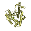
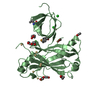
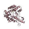
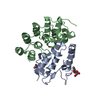
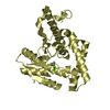
- PDB-6uej: Crystal structure of human zinc finger antiviral protein bound to RNA -
+ Open data

- Basic information
| Entry | Database: PDB / ID: 6uej | ||||||
|---|---|---|---|---|---|---|---|
| Title | Crystal structure of human zinc finger antiviral protein bound to RNA | ||||||
 Components Components |
| ||||||
 Keywords Keywords |  ANTIVIRAL PROTEIN / Zinc Finger Antiviral protein / ZAP / RNA binding domain ANTIVIRAL PROTEIN / Zinc Finger Antiviral protein / ZAP / RNA binding domain | ||||||
| Function / homology |  Function and homology information Function and homology informationpositive regulation of RIG-I signaling pathway / positive regulation of mRNA catabolic process / negative regulation of viral genome replication / positive regulation of interferon-alpha production / positive regulation of type I interferon production / positive regulation of interferon-beta production / response to virus / Signaling by BRAF and RAF1 fusions / positive regulation of canonical NF-kappaB signal transduction / defense response to virus ...positive regulation of RIG-I signaling pathway / positive regulation of mRNA catabolic process / negative regulation of viral genome replication / positive regulation of interferon-alpha production / positive regulation of type I interferon production / positive regulation of interferon-beta production / response to virus / Signaling by BRAF and RAF1 fusions / positive regulation of canonical NF-kappaB signal transduction / defense response to virus / cadherin binding / innate immune response / RNA binding / metal ion binding / nucleus / cytosol / cytoplasmSimilarity search - Function | ||||||
| Biological species |  Homo sapiens (human) Homo sapiens (human)synthetic construct (others) | ||||||
| Method | X-RAY DIFFRACTION / SYNCHROTRON / MOLECULAR REPLACEMENT / Resolution: 2.21 Å | ||||||
 Authors Authors | Meagher, J.L. / Smith, J.L. | ||||||
| Funding support |  United States, 1items United States, 1items
| ||||||
 Citation Citation | Journal: Proc.Natl.Acad.Sci.USA / Year: 2019 Title: Structure of the zinc-finger antiviral protein in complex with RNA reveals a mechanism for selective targeting of CG-rich viral sequences. Authors: Meagher, J.L. / Takata, M. / Goncalves-Carneiro, D. / Keane, S.C. / Rebendenne, A. / Ong, H. / Orr, V.K. / MacDonald, M.R. / Stuckey, J.A. / Bieniasz, P.D. / Smith, J.L. | ||||||
| History |
|
- Structure visualization
| Structure viewer | Molecule: MolmilJmol/JSmol |
|---|
- Downloads & links
-Download
| PDBx/mmCIF format | 6uej.cif.gz | 64.6 KB | Display | PDBx/mmCIF format |
|---|---|---|---|---|
| PDB format | pdb6uej.ent.gz | 42.9 KB | Display | PDB format |
| PDBx/mmJSON format | 6uej.json.gz | Tree view | PDBx/mmJSON format | |
| Others |  Other downloads Other downloads |
-Validation report
| Arichive directory | https://data.pdbj.org/pub/pdb/validation_reports/ue/6uejftp://data.pdbj.org/pub/pdb/validation_reports/ue/6uej | HTTPS FTP |
|---|
-Related structure data
| Related structure data |  6ueiC  3u9gS S: Starting model for refinement C: citing same article ( |
|---|---|
| Similar structure data |


-Links
 PDBj
PDBj


- Assembly
| Deposited unit | 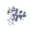
| ||||||||
|---|---|---|---|---|---|---|---|---|---|
| 1 |
| ||||||||
| Unit cell |
|
-Components
| #1: Protein | Mass: 25991.076 Da / Num. of mol.: 1 Source method: isolated from a genetically manipulated source Source: (gene. exp.) Homo sapiens (human) / Gene: ZC3HAV1, ZC3HDC2, PRO1677 / Production host:  Escherichia coli (E. coli) / References: UniProt: Q7Z2W4 Escherichia coli (E. coli) / References: UniProt: Q7Z2W4 | ||||||
|---|---|---|---|---|---|---|---|
| #2: RNA chain | Mass: 911.596 Da / Num. of mol.: 1 / Source method: obtained synthetically / Source: (synth.) synthetic construct (others) | ||||||
| #3: Chemical | ChemComp-ZN /   Mass: 65.409 Da / Num. of mol.: 4 / Source method: obtained synthetically / Formula: Zn Mass: 65.409 Da / Num. of mol.: 4 / Source method: obtained synthetically / Formula: Zn#4: Chemical | ChemComp-SPM / | Spermine  Mass: 202.340 Da / Num. of mol.: 1 / Source method: obtained synthetically / Formula: C10H26N4 Mass: 202.340 Da / Num. of mol.: 1 / Source method: obtained synthetically / Formula: C10H26N4#5: Water | ChemComp-HOH / | Water Mass: 18.015 Da / Num. of mol.: 93 / Source method: isolated from a natural source / Formula: H2O Mass: 18.015 Da / Num. of mol.: 93 / Source method: isolated from a natural source / Formula: H2OHas ligand of interest | N | |
-Experimental details
-Experiment
| Experiment | Method: X-RAY DIFFRACTION / Number of used crystals: 1 |
|---|
- Sample preparation
| Crystal | Density Matthews: 3.03 Å3/Da / Density % sol: 59.4 % |
|---|---|
| Crystal grow | Temperature: 293.15 K / Method: vapor diffusion, sitting drop / pH: 7.5 Details: 7% Peg 8000, 30mM magnesium chloride, 10mM spermine, 50mM HEPES pH 7.5 |
-Data collection
| Diffraction | Mean temperature: 100 K / Serial crystal experiment: N | ||||||||||||||||||||||||||||||||||||||||||||||||||||||||||||||||||||||||||||||||||||||||||||||||||||
|---|---|---|---|---|---|---|---|---|---|---|---|---|---|---|---|---|---|---|---|---|---|---|---|---|---|---|---|---|---|---|---|---|---|---|---|---|---|---|---|---|---|---|---|---|---|---|---|---|---|---|---|---|---|---|---|---|---|---|---|---|---|---|---|---|---|---|---|---|---|---|---|---|---|---|---|---|---|---|---|---|---|---|---|---|---|---|---|---|---|---|---|---|---|---|---|---|---|---|---|---|---|
| Diffraction source | Source: SYNCHROTRON / Site: APS / Beamline: 21-ID-D / Wavelength: 0.9762 Å | ||||||||||||||||||||||||||||||||||||||||||||||||||||||||||||||||||||||||||||||||||||||||||||||||||||
| Detector | Type: DECTRIS EIGER X 9M / Detector: PIXEL / Date: Feb 7, 2019 | ||||||||||||||||||||||||||||||||||||||||||||||||||||||||||||||||||||||||||||||||||||||||||||||||||||
| Radiation | Protocol: SINGLE WAVELENGTH / Monochromatic (M) / Laue (L): M / Scattering type: x-ray | ||||||||||||||||||||||||||||||||||||||||||||||||||||||||||||||||||||||||||||||||||||||||||||||||||||
| Radiation wavelength | Wavelength: 0.9762 Å / Relative weight: 1 | ||||||||||||||||||||||||||||||||||||||||||||||||||||||||||||||||||||||||||||||||||||||||||||||||||||
| Reflection | Resolution: 2.2→40.668 Å / Num. obs: 18081 / % possible obs: 99.8 % / Redundancy: 20.564 % / Biso Wilson estimate: 64.21 Å2 / CC1/2: 0.999 / Rmerge(I) obs: 0.079 / Rrim(I) all: 0.081 / Χ2: 1.087 / Net I/σ(I): 20.69 / Num. measured all: 371826 | ||||||||||||||||||||||||||||||||||||||||||||||||||||||||||||||||||||||||||||||||||||||||||||||||||||
| Reflection shell | Diffraction-ID: 1
|
- Processing
| Software |
| ||||||||||||||||||||||||||||||||||||||||||||||||||||||||||||||||||||||||||||||||||||||||||||||||||||||||||||
|---|---|---|---|---|---|---|---|---|---|---|---|---|---|---|---|---|---|---|---|---|---|---|---|---|---|---|---|---|---|---|---|---|---|---|---|---|---|---|---|---|---|---|---|---|---|---|---|---|---|---|---|---|---|---|---|---|---|---|---|---|---|---|---|---|---|---|---|---|---|---|---|---|---|---|---|---|---|---|---|---|---|---|---|---|---|---|---|---|---|---|---|---|---|---|---|---|---|---|---|---|---|---|---|---|---|---|---|---|---|
| Refinement | Method to determine structure: MOLECULAR REPLACEMENT Starting model: 3U9G Resolution: 2.21→38 Å / Cor.coef. Fo:Fc: 0.934 / Cor.coef. Fo:Fc free: 0.919 / SU R Cruickshank DPI: 0.19 / Cross valid method: THROUGHOUT / σ(F): 0 / SU R Blow DPI: 0.196 / SU Rfree Blow DPI: 0.18 / SU Rfree Cruickshank DPI: 0.177
| ||||||||||||||||||||||||||||||||||||||||||||||||||||||||||||||||||||||||||||||||||||||||||||||||||||||||||||
| Displacement parameters | Biso max: 167.22 Å2 / Biso mean: 75.67 Å2 / Biso min: 43.74 Å2
| ||||||||||||||||||||||||||||||||||||||||||||||||||||||||||||||||||||||||||||||||||||||||||||||||||||||||||||
| Refine analyze | Luzzati coordinate error obs: 0.32 Å | ||||||||||||||||||||||||||||||||||||||||||||||||||||||||||||||||||||||||||||||||||||||||||||||||||||||||||||
| Refinement step | Cycle: final / Resolution: 2.21→38 Å
| ||||||||||||||||||||||||||||||||||||||||||||||||||||||||||||||||||||||||||||||||||||||||||||||||||||||||||||
| Refine LS restraints |
| ||||||||||||||||||||||||||||||||||||||||||||||||||||||||||||||||||||||||||||||||||||||||||||||||||||||||||||
| LS refinement shell | Resolution: 2.21→2.22 Å / Rfactor Rfree error: 0 / Total num. of bins used: 47
|
