 Movie
Movie Controller


+ Open data

- Basic information
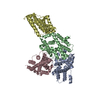
| Entry | Database: PDB / ID: 6u1n | |||||||||
|---|---|---|---|---|---|---|---|---|---|---|
| Title | GPCR-Beta arrestin structure in lipid bilayer | |||||||||
 Components Components |
| |||||||||
 Keywords Keywords |  SIGNALING PROTEIN/IMMUNE SYSTEM / Arrestin / GPCR / complex / signaling / SIGNALING PROTEIN-IMMUNE SYSTEM complex SIGNALING PROTEIN/IMMUNE SYSTEM / Arrestin / GPCR / complex / signaling / SIGNALING PROTEIN-IMMUNE SYSTEM complex | |||||||||
| Function / homology |  Function and homology informationV2 vasopressin receptor binding / alpha-1A adrenergic receptor binding / follicle-stimulating hormone receptor binding / Activation of SMO / sensory perception of touch / renal water retention / Defective AVP does not bind AVPR2 and causes neurohypophyseal diabetes insipidus (NDI) / G alpha (s) signalling events / Vasopressin-like receptors / regulation of systemic arterial blood pressure by vasopressin ...V2 vasopressin receptor binding / alpha-1A adrenergic receptor binding / follicle-stimulating hormone receptor binding / Activation of SMO / sensory perception of touch / renal water retention / Defective AVP does not bind AVPR2 and causes neurohypophyseal diabetes insipidus (NDI) / G alpha (s) signalling events / Vasopressin-like receptors / regulation of systemic arterial blood pressure by vasopressin / vasopressin receptor activity / alpha-1B adrenergic receptor binding / follicle-stimulating hormone signaling pathway / protein phosphorylated amino acid binding / angiotensin receptor binding / Lysosome Vesicle Biogenesis / AP-2 adaptor complex binding / Muscarinic acetylcholine receptors / symmetric synapse / Golgi Associated Vesicle Biogenesis / phospholipase C-activating G protein-coupled acetylcholine receptor signaling pathway / MAP2K and MAPK activation / Ub-specific processing proteases / G protein-coupled acetylcholine receptor activity / regulation of smooth muscle contraction / positive regulation of systemic arterial blood pressure / cholinergic synapse / hemostasis / telencephalon development / positive regulation of smooth muscle cell apoptotic process / adenylate cyclase-inhibiting G protein-coupled acetylcholine receptor signaling pathway / negative regulation of interleukin-8 production / Cargo recognition for clathrin-mediated endocytosis / Clathrin-mediated endocytosis / clathrin adaptor activity / regulation of G protein-coupled receptor signaling pathway / G protein-coupled serotonin receptor activity / arrestin family protein binding / G protein-coupled receptor internalization / Thrombin signalling through proteinase activated receptors (PARs) / regulation of heart contraction / positive regulation of intracellular signal transduction / mitogen-activated protein kinase kinase binding / positive regulation of Rho protein signal transduction / clathrin binding / stress fiber assembly / negative regulation of Notch signaling pathway / immunoglobulin complex / pseudopodium / positive regulation of insulin secretion involved in cellular response to glucose stimulus / G protein-coupled receptor signaling pathway, coupled to cyclic nucleotide second messenger / cysteine-type endopeptidase inhibitor activity involved in apoptotic process / negative regulation of interleukin-6 production / positive regulation of receptor internalization / phototransduction / endocytic vesicle / asymmetric synapse / axon terminus / clathrin-coated pit / positive regulation of vasoconstriction / negative regulation of protein ubiquitination / cellular response to hormone stimulus / insulin-like growth factor receptor binding / activation of adenylate cyclase activity / visual perception / presynaptic modulation of chemical synaptic transmission / GTPase activator activity / negative regulation of protein phosphorylation / positive regulation of protein ubiquitination / response to cytokine / G protein-coupled receptor binding / nuclear estrogen receptor binding / peptide binding / phosphoprotein binding / clathrin-coated endocytic vesicle membrane / response to virus / adenylate cyclase-modulating G protein-coupled receptor signaling pathway / G protein-coupled acetylcholine receptor signaling pathway / negative regulation of ERK1 and ERK2 cascade / Vasopressin regulates renal water homeostasis via Aquaporins / endocytosis / Cargo recognition for clathrin-mediated endocytosis / protein transport / presynaptic membrane / Clathrin-mediated endocytosis / positive regulation of peptidyl-serine phosphorylation / nervous system development / G alpha (i) signalling events / ubiquitin-dependent protein catabolic process / cytoplasmic vesicle / G alpha (s) signalling events / chemical synaptic transmission / postsynaptic membrane / basolateral plasma membrane / proteasome-mediated ubiquitin-dependent protein catabolic process / regulation of apoptotic process / negative regulation of neuron apoptotic process / transmembrane transporter binding / dendritic spine / positive regulation of MAPK cascade Function and homology informationV2 vasopressin receptor binding / alpha-1A adrenergic receptor binding / follicle-stimulating hormone receptor binding / Activation of SMO / sensory perception of touch / renal water retention / Defective AVP does not bind AVPR2 and causes neurohypophyseal diabetes insipidus (NDI) / G alpha (s) signalling events / Vasopressin-like receptors / regulation of systemic arterial blood pressure by vasopressin ...V2 vasopressin receptor binding / alpha-1A adrenergic receptor binding / follicle-stimulating hormone receptor binding / Activation of SMO / sensory perception of touch / renal water retention / Defective AVP does not bind AVPR2 and causes neurohypophyseal diabetes insipidus (NDI) / G alpha (s) signalling events / Vasopressin-like receptors / regulation of systemic arterial blood pressure by vasopressin / vasopressin receptor activity / alpha-1B adrenergic receptor binding / follicle-stimulating hormone signaling pathway / protein phosphorylated amino acid binding / angiotensin receptor binding / Lysosome Vesicle Biogenesis / AP-2 adaptor complex binding / Muscarinic acetylcholine receptors / symmetric synapse / Golgi Associated Vesicle Biogenesis / phospholipase C-activating G protein-coupled acetylcholine receptor signaling pathway / MAP2K and MAPK activation / Ub-specific processing proteases / G protein-coupled acetylcholine receptor activity / regulation of smooth muscle contraction / positive regulation of systemic arterial blood pressure / cholinergic synapse / hemostasis / telencephalon development / positive regulation of smooth muscle cell apoptotic process / adenylate cyclase-inhibiting G protein-coupled acetylcholine receptor signaling pathway / negative regulation of interleukin-8 production / Cargo recognition for clathrin-mediated endocytosis / Clathrin-mediated endocytosis / clathrin adaptor activity / regulation of G protein-coupled receptor signaling pathway / G protein-coupled serotonin receptor activity / arrestin family protein binding / G protein-coupled receptor internalization / Thrombin signalling through proteinase activated receptors (PARs) / regulation of heart contraction / positive regulation of intracellular signal transduction / mitogen-activated protein kinase kinase binding / positive regulation of Rho protein signal transduction / clathrin binding / stress fiber assembly / negative regulation of Notch signaling pathway / immunoglobulin complex / pseudopodium / positive regulation of insulin secretion involved in cellular response to glucose stimulus / G protein-coupled receptor signaling pathway, coupled to cyclic nucleotide second messenger / cysteine-type endopeptidase inhibitor activity involved in apoptotic process / negative regulation of interleukin-6 production / positive regulation of receptor internalization / phototransduction / endocytic vesicle / asymmetric synapse / axon terminus / clathrin-coated pit / positive regulation of vasoconstriction / negative regulation of protein ubiquitination / cellular response to hormone stimulus / insulin-like growth factor receptor binding / activation of adenylate cyclase activity / visual perception / presynaptic modulation of chemical synaptic transmission / GTPase activator activity / negative regulation of protein phosphorylation / positive regulation of protein ubiquitination / response to cytokine / G protein-coupled receptor binding / nuclear estrogen receptor binding / peptide binding / phosphoprotein binding / clathrin-coated endocytic vesicle membrane / response to virus / adenylate cyclase-modulating G protein-coupled receptor signaling pathway / G protein-coupled acetylcholine receptor signaling pathway / negative regulation of ERK1 and ERK2 cascade / Vasopressin regulates renal water homeostasis via Aquaporins / endocytosis / Cargo recognition for clathrin-mediated endocytosis / protein transport / presynaptic membrane / Clathrin-mediated endocytosis / positive regulation of peptidyl-serine phosphorylation / nervous system development / G alpha (i) signalling events / ubiquitin-dependent protein catabolic process / cytoplasmic vesicle / G alpha (s) signalling events / chemical synaptic transmission / postsynaptic membrane / basolateral plasma membrane / proteasome-mediated ubiquitin-dependent protein catabolic process / regulation of apoptotic process / negative regulation of neuron apoptotic process / transmembrane transporter binding / dendritic spine / positive regulation of MAPK cascadeSimilarity search - Function | |||||||||
| Biological species |  Homo sapiens (human) Homo sapiens (human) Rattus norvegicus (Norway rat) Rattus norvegicus (Norway rat) | |||||||||
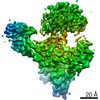
| Method | ELECTRON MICROSCOPY / single particle reconstruction / cryo EM / Resolution: 4 Å | |||||||||
 Authors Authors | Staus, D.P. / Hu, H. / Robertson, M.J. / Kleinhenz, A.L.W. / Wingler, L.M. / Capel, W.D. / Latorraca, N.R. / Lefkowitz, R.J. / Skiniotis, G. | |||||||||
| Funding support |  United States, 2items United States, 2items
| |||||||||
 Citation Citation | Journal: Nature / Year: 2020 Title: Structure of the M2 muscarinic receptor-β-arrestin complex in a lipid nanodisc. Authors: Dean P Staus / Hongli Hu / Michael J Robertson / Alissa L W Kleinhenz / Laura M Wingler / William D Capel / Naomi R Latorraca / Robert J Lefkowitz / Georgios Skiniotis /  Abstract: After activation by an agonist, G-protein-coupled receptors (GPCRs) recruit β-arrestin, which desensitizes heterotrimeric G-protein signalling and promotes receptor endocytosis. Additionally, β- ...After activation by an agonist, G-protein-coupled receptors (GPCRs) recruit β-arrestin, which desensitizes heterotrimeric G-protein signalling and promotes receptor endocytosis. Additionally, β-arrestin directly regulates many cell signalling pathways that can induce cellular responses distinct from that of G proteins. In contrast to G proteins, for which there are many high-resolution structures in complex with GPCRs, the molecular mechanisms underlying the interaction of β-arrestin with GPCRs are much less understood. Here we present a cryo-electron microscopy structure of β-arrestin 1 (βarr1) in complex with M2 muscarinic receptor (M2R) reconstituted in lipid nanodiscs. The M2R-βarr1 complex displays a multimodal network of flexible interactions, including binding of the N domain of βarr1 to phosphorylated receptor residues and insertion of the finger loop of βarr1 into the M2R seven-transmembrane bundle, which adopts a conformation similar to that in the M2R-heterotrimeric G protein complex. Moreover, the cryo-electron microscopy map reveals that the C-edge of βarr1 engages the lipid bilayer. Through atomistic simulations and biophysical, biochemical and cellular assays, we show that the C-edge is critical for stable complex formation, βarr1 recruitment, receptor internalization, and desensitization of G-protein activation. Taken together, these data suggest that the cooperative interactions of β-arrestin with both the receptor and the phospholipid bilayer contribute to its functional versatility. | |||||||||
| History |
|
- Structure visualization
| Movie |
Movie viewer |
|---|---|
| Structure viewer | Molecule: MolmilJmol/JSmol |
- Downloads & links
-Download
| PDBx/mmCIF format | 6u1n.cif.gz | 163.5 KB | Display | PDBx/mmCIF format |
|---|---|---|---|---|
| PDB format | pdb6u1n.ent.gz | 119.8 KB | Display | PDB format |
| PDBx/mmJSON format | 6u1n.json.gz | Tree view | PDBx/mmJSON format | |
| Others |  Other downloads Other downloads |
-Validation report
| Arichive directory | https://data.pdbj.org/pub/pdb/validation_reports/u1/6u1nftp://data.pdbj.org/pub/pdb/validation_reports/u1/6u1n | HTTPS FTP |
|---|
-Related structure data
| Related structure data | 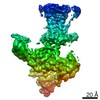 20612MC M: map data used to model this data C: citing same article ( |
|---|---|
| Similar structure data |


-Links
 PDBj
PDBj


- Assembly
| Deposited unit | 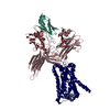
|
|---|---|
| 1 |
|
-Components
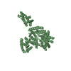
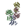
| #1: Protein | Mass: 56616.176 Da / Num. of mol.: 1 / Fragment: M2 UNP residues 2-466 + V2 UNP residues 343-371 Source method: isolated from a genetically manipulated source Source: (gene. exp.) Homo sapiens (human) / Gene: CHRM2, AVPR2, ADHR, DIR, DIR3, V2R / Production host: Homo sapiens (human) / References: UniProt: P08172, UniProt: P30518 |
|---|---|
| #2: Protein | Arrestin / Arrestin beta-1 Mass: 45017.180 Da / Num. of mol.: 1 Source method: isolated from a genetically manipulated source Source: (gene. exp.) Rattus norvegicus (Norway rat) / Gene: Arrb1 / Production host:  Escherichia coli (E. coli) / References: UniProt: P29066 Escherichia coli (E. coli) / References: UniProt: P29066 |
| #3: Antibody | Mass: 25512.354 Da / Num. of mol.: 1 Source method: isolated from a genetically manipulated source Source: (gene. exp.) Homo sapiens (human) / Production host: Escherichia coli (E. coli) / References: UniProt: V9HW68 |
| #4: Antibody | Mass: 23435.064 Da / Num. of mol.: 1 Source method: isolated from a genetically manipulated source Source: (gene. exp.) Homo sapiens (human) / Production host: Escherichia coli (E. coli) / References: UniProt: Q7Z3Y4 |
| #5: Chemical | ChemComp-2CU /   Mass: 437.944 Da / Num. of mol.: 1 / Source method: obtained synthetically / Formula: C19H24ClN5O3S Mass: 437.944 Da / Num. of mol.: 1 / Source method: obtained synthetically / Formula: C19H24ClN5O3S |
| Has ligand of interest | N |
-Experimental details
-Experiment
| Experiment | Method: ELECTRON MICROSCOPY |
|---|---|
| EM experiment | Aggregation state: PARTICLE / 3D reconstruction method: single particle reconstruction |
- Sample preparation
| Component |
| |||||||||||||||||||||||||||||||||||
|---|---|---|---|---|---|---|---|---|---|---|---|---|---|---|---|---|---|---|---|---|---|---|---|---|---|---|---|---|---|---|---|---|---|---|---|---|
| Source (natural) |
| |||||||||||||||||||||||||||||||||||
| Source (recombinant) |
| |||||||||||||||||||||||||||||||||||
| Buffer solution | pH: 7.4 | |||||||||||||||||||||||||||||||||||
| Specimen | Conc.: 2 mg/ml / Embedding applied: NO / Shadowing applied: NO / Staining applied: NO / Vitrification applied: YES | |||||||||||||||||||||||||||||||||||
| Specimen support | Details: unspecified | |||||||||||||||||||||||||||||||||||
| Vitrification | Cryogen name: ETHANE |
- Electron microscopy imaging
| Experimental equipment |  Model: Titan Krios / Image courtesy: FEI Company |
|---|---|
| Microscopy | Model: FEI TITAN KRIOS |
| Electron gun | Electron source: FIELD EMISSION GUN / Accelerating voltage: 300 kV / Illumination mode: FLOOD BEAM |
| Electron lens | Mode: BRIGHT FIELDBright-field microscopy / Nominal magnification: 47169 X / Calibrated magnification: 47169 X / Nominal defocus max: 2200 nm / Nominal defocus min: 1200 nm / C2 aperture diameter: 50 µm |
| Specimen holder | Cryogen: NITROGEN / Specimen holder model: FEI TITAN KRIOS AUTOGRID HOLDER |
| Image recording | Electron dose: 50 e/Å2 / Detector mode: COUNTING / Film or detector model: GATAN K2 QUANTUM (4k x 4k) |
- Processing
| Software | Name: PHENIX / Version: 1.15rc3_3435: / Classification: refinement | ||||||||||||||||||||||||||||||||
|---|---|---|---|---|---|---|---|---|---|---|---|---|---|---|---|---|---|---|---|---|---|---|---|---|---|---|---|---|---|---|---|---|---|
| EM software |
| ||||||||||||||||||||||||||||||||
| CTF correction | Type: PHASE FLIPPING AND AMPLITUDE CORRECTION | ||||||||||||||||||||||||||||||||
| Particle selection | Num. of particles selected: 11700000 | ||||||||||||||||||||||||||||||||
| Symmetry | Point symmetry: C1 (asymmetric) | ||||||||||||||||||||||||||||||||
| 3D reconstruction | Resolution: 4 Å / Resolution method: FSC 0.143 CUT-OFF / Num. of particles: 145618 / Symmetry type: POINT | ||||||||||||||||||||||||||||||||
| Atomic model building | Protocol: FLEXIBLE FIT / Space: REAL | ||||||||||||||||||||||||||||||||
| Refine LS restraints |
|
