National Institutes of Health/National Heart, Lung, and Blood Institute (NIH/NHLBI)
HL16037
United States
National Institutes of Health/National Institute of Neurological Disorders and Stroke (NIH/NINDS)
NS092695
United States
Citation
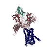
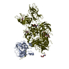
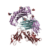
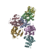
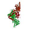
Journal: Nature / Year: 2020 Title: Structure of the M2 muscarinic receptor-β-arrestin complex in a lipid nanodisc. Authors: Dean P Staus / Hongli Hu / Michael J Robertson / Alissa L W Kleinhenz / Laura M Wingler / William D Capel / Naomi R Latorraca / Robert J Lefkowitz / Georgios Skiniotis / Abstract: After activation by an agonist, G-protein-coupled receptors (GPCRs) recruit β-arrestin, which desensitizes heterotrimeric G-protein signalling and promotes receptor endocytosis. Additionally, β- ...After activation by an agonist, G-protein-coupled receptors (GPCRs) recruit β-arrestin, which desensitizes heterotrimeric G-protein signalling and promotes receptor endocytosis. Additionally, β-arrestin directly regulates many cell signalling pathways that can induce cellular responses distinct from that of G proteins. In contrast to G proteins, for which there are many high-resolution structures in complex with GPCRs, the molecular mechanisms underlying the interaction of β-arrestin with GPCRs are much less understood. Here we present a cryo-electron microscopy structure of β-arrestin 1 (βarr1) in complex with M2 muscarinic receptor (M2R) reconstituted in lipid nanodiscs. The M2R-βarr1 complex displays a multimodal network of flexible interactions, including binding of the N domain of βarr1 to phosphorylated receptor residues and insertion of the finger loop of βarr1 into the M2R seven-transmembrane bundle, which adopts a conformation similar to that in the M2R-heterotrimeric G protein complex. Moreover, the cryo-electron microscopy map reveals that the C-edge of βarr1 engages the lipid bilayer. Through atomistic simulations and biophysical, biochemical and cellular assays, we show that the C-edge is critical for stable complex formation, βarr1 recruitment, receptor internalization, and desensitization of G-protein activation. Taken together, these data suggest that the cooperative interactions of β-arrestin with both the receptor and the phospholipid bilayer contribute to its functional versatility.
History
Deposition
Aug 16, 2019
-
Header (metadata) release
Oct 2, 2019
-
Map release
Feb 26, 2020
-
Update
Mar 25, 2020
-
Current status
Mar 25, 2020
Processing site: RCSB / Status: Released
-
Structure visualization
Movie
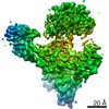
Surface view with section colored by density value
Supramolecule #1: Phosphorylated human muscarinic acetylcholine receptor M2 in comp...
Supramolecule
Name: Phosphorylated human muscarinic acetylcholine receptor M2 in complex with rat beta-arrestin1, stabilized by an antibody fragment (Fab30). type: complex / ID: 1 / Parent: 0 / Macromolecule list: #1-#4
-
Supramolecule #2: Phosphorylated human muscarinic acetylcholine receptor M2
Supramolecule
Name: Phosphorylated human muscarinic acetylcholine receptor M2 type: complex / ID: 2 / Parent: 1 / Macromolecule list: #1 Details: Phosphorylated M2R was generated by ligating a synthetic phosphopeptide derived from the vasopressin-2-receptor (V2Rpp) using the enzyme sortase.
Source (natural)
Organism: Homo sapiens (human)
Recombinant expression
Organism: Homo sapiens (human)
-
Supramolecule #3: Cysteine-free rat beta-arrestin 1 truncated at amino acid 393
In the structure databanks used in Yorodumi, some data are registered as the other names, "COVID-19 virus" and "2019-nCoV". Here are the details of the virus and the list of structure data.
Jan 31, 2019. EMDB accession codes are about to change! (news from PDBe EMDB page)
EMDB accession codes are about to change! (news from PDBe EMDB page)
The allocation of 4 digits for EMDB accession codes will soon come to an end. Whilst these codes will remain in use, new EMDB accession codes will include an additional digit and will expand incrementally as the available range of codes is exhausted. The current 4-digit format prefixed with “EMD-” (i.e. EMD-XXXX) will advance to a 5-digit format (i.e. EMD-XXXXX), and so on. It is currently estimated that the 4-digit codes will be depleted around Spring 2019, at which point the 5-digit format will come into force.
The EM Navigator/Yorodumi systems omit the EMD- prefix.
Related info.:Q: What is EMD? / ID/Accession-code notation in Yorodumi/EM Navigator
Yorodumi is a browser for structure data from EMDB, PDB, SASBDB, etc.
This page is also the successor to EM Navigator detail page, and also detail information page/front-end page for Omokage search.
The word "yorodu" (or yorozu) is an old Japanese word meaning "ten thousand". "mi" (miru) is to see.
Related info.:EMDB / PDB / SASBDB / Comparison of 3 databanks / Yorodumi Search / Aug 31, 2016. New EM Navigator & Yorodumi / Yorodumi Papers / Jmol/JSmol / Function and homology information / Changes in new EM Navigator and Yorodumi
 Movie
Movie Controller


 Open data
 Basic information
 Map data
 Sample
 Function and homology information
 V2 vasopressin receptor binding /

 Authors
 United States, 2 items
  Citation
 Structure visualization
 Downloads & links
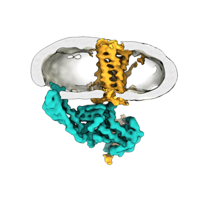 emd_20612.png
 http://ftp.pdbj.org/pub/emdb/structures/EMD-20612


 Sample components

 Processing
 Electron microscopy
