Movie
Movie Controller
 Structure viewers
 About Yorodumi Papers


+Search query
-Structure paper
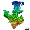
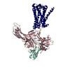
| Title | Structure of the M2 muscarinic receptor-β-arrestin complex in a lipid nanodisc. |
|---|---|
| Journal, issue, pages | Nature, Vol. 579, Issue 7798, Page 297-302, Year 2020 |
| Publish date | Jan 16, 2020 |
 Authors Authors | Dean P Staus / Hongli Hu / Michael J Robertson / Alissa L W Kleinhenz / Laura M Wingler / William D Capel / Naomi R Latorraca / Robert J Lefkowitz / Georgios Skiniotis /   |
| PubMed Abstract | After activation by an agonist, G-protein-coupled receptors (GPCRs) recruit β-arrestin, which desensitizes heterotrimeric G-protein signalling and promotes receptor endocytosis. Additionally, β- ...After activation by an agonist, G-protein-coupled receptors (GPCRs) recruit β-arrestin, which desensitizes heterotrimeric G-protein signalling and promotes receptor endocytosis. Additionally, β-arrestin directly regulates many cell signalling pathways that can induce cellular responses distinct from that of G proteins. In contrast to G proteins, for which there are many high-resolution structures in complex with GPCRs, the molecular mechanisms underlying the interaction of β-arrestin with GPCRs are much less understood. Here we present a cryo-electron microscopy structure of β-arrestin 1 (βarr1) in complex with M2 muscarinic receptor (M2R) reconstituted in lipid nanodiscs. The M2R-βarr1 complex displays a multimodal network of flexible interactions, including binding of the N domain of βarr1 to phosphorylated receptor residues and insertion of the finger loop of βarr1 into the M2R seven-transmembrane bundle, which adopts a conformation similar to that in the M2R-heterotrimeric G protein complex. Moreover, the cryo-electron microscopy map reveals that the C-edge of βarr1 engages the lipid bilayer. Through atomistic simulations and biophysical, biochemical and cellular assays, we show that the C-edge is critical for stable complex formation, βarr1 recruitment, receptor internalization, and desensitization of G-protein activation. Taken together, these data suggest that the cooperative interactions of β-arrestin with both the receptor and the phospholipid bilayer contribute to its functional versatility. |
 External links External links | Nature / PubMed:31945772 / PubMed Central |
| Methods | EM (single particle) |
| Resolution | 3.6 - 4.0 Å |
| Structure data | EMDB-20612, PDB-6u1n: 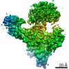 EMDB-20948: |
| Chemicals |  ChemComp-2CU: |
| Source |
|
 Keywords Keywords |  SIGNALING PROTEIN/IMMUNE SYSTEM / Arrestin / GPCR / complex / signaling / SIGNALING PROTEIN-IMMUNE SYSTEM complex SIGNALING PROTEIN/IMMUNE SYSTEM / Arrestin / GPCR / complex / signaling / SIGNALING PROTEIN-IMMUNE SYSTEM complex |