 Movie
Movie Controller


+ Open data

- Basic information
| Entry | Database: PDB / ID: 6trj | ||||||
|---|---|---|---|---|---|---|---|
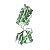
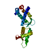
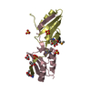
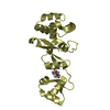
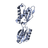
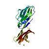
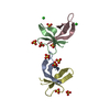
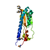
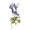
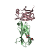
| Title | LEDGF/p75 IBD dimer | ||||||
 Components Components | PC4 and SFRS1-interacting protein | ||||||
 Keywords Keywords |  PROTEIN BINDING / protein-protein interaction / transcription factor PROTEIN BINDING / protein-protein interaction / transcription factor | ||||||
| Function / homology |  Function and homology informationsupercoiled DNA binding / Integration of viral DNA into host genomic DNA / Autointegration results in viral DNA circles / 2-LTR circle formation / Formation of WDR5-containing histone-modifying complexes / Vpr-mediated nuclear import of PICs / Integration of provirus / APOBEC3G mediated resistance to HIV-1 infection / mRNA 5'-splice site recognition / heterochromatin ...supercoiled DNA binding / Integration of viral DNA into host genomic DNA / Autointegration results in viral DNA circles / 2-LTR circle formation / Formation of WDR5-containing histone-modifying complexes / Vpr-mediated nuclear import of PICs / Integration of provirus / APOBEC3G mediated resistance to HIV-1 infection / mRNA 5'-splice site recognition / heterochromatin / nuclear periphery / euchromatin / response to heat / DNA-binding transcription factor binding / response to oxidative stress / transcription coactivator activity / chromatin remodeling / chromatin binding / positive regulation of transcription by RNA polymerase II / RNA binding / nucleoplasm / nucleus / cytosol Function and homology informationsupercoiled DNA binding / Integration of viral DNA into host genomic DNA / Autointegration results in viral DNA circles / 2-LTR circle formation / Formation of WDR5-containing histone-modifying complexes / Vpr-mediated nuclear import of PICs / Integration of provirus / APOBEC3G mediated resistance to HIV-1 infection / mRNA 5'-splice site recognition / heterochromatin ...supercoiled DNA binding / Integration of viral DNA into host genomic DNA / Autointegration results in viral DNA circles / 2-LTR circle formation / Formation of WDR5-containing histone-modifying complexes / Vpr-mediated nuclear import of PICs / Integration of provirus / APOBEC3G mediated resistance to HIV-1 infection / mRNA 5'-splice site recognition / heterochromatin / nuclear periphery / euchromatin / response to heat / DNA-binding transcription factor binding / response to oxidative stress / transcription coactivator activity / chromatin remodeling / chromatin binding / positive regulation of transcription by RNA polymerase II / RNA binding / nucleoplasm / nucleus / cytosolSimilarity search - Function | ||||||
| Biological species |  Homo sapiens (human) Homo sapiens (human) | ||||||
| Method | X-RAY DIFFRACTION / AB INITIO PHASING / Resolution: 1.3 Å | ||||||
 Authors Authors | Kugler, M. / Brynda, J. | ||||||
 Citation Citation | Journal: Structure Title: Fine-tuning of the LEDGF/p75 interaction network by dimerization Authors: Lux, V. / Brouns, T. / Cermakova, K. / Srb, P. / Fabry, M. / Madlikova, M. / Horejsi, M. / Kugler, M. / Brynda, J. / Novak, P. / DeRijck, J. / Christ, F. / Debyser, Z. / Veverka, V. | ||||||
| History |
|
- Structure visualization
| Structure viewer | Molecule: MolmilJmol/JSmol |
|---|
- Downloads & links
-Download
| PDBx/mmCIF format | 6trj.cif.gz | 53.6 KB | Display | PDBx/mmCIF format |
|---|---|---|---|---|
| PDB format | pdb6trj.ent.gz | 38.1 KB | Display | PDB format |
| PDBx/mmJSON format | 6trj.json.gz | Tree view | PDBx/mmJSON format | |
| Others |  Other downloads Other downloads |
-Validation report
| Arichive directory | https://data.pdbj.org/pub/pdb/validation_reports/tr/6trjftp://data.pdbj.org/pub/pdb/validation_reports/tr/6trj | HTTPS FTP |
|---|
-Related structure data
| Similar structure data |
|---|
-Links
 PDBj
PDBj


- Assembly
| Deposited unit | 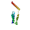
| |||||||||
|---|---|---|---|---|---|---|---|---|---|---|
| 1 | 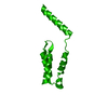
| |||||||||
| Unit cell |
| |||||||||
| Components on special symmetry positions |
|
-Components
| #1: Protein | Mass: 10519.277 Da / Num. of mol.: 1 Source method: isolated from a genetically manipulated source Source: (gene. exp.) Homo sapiens (human) / Gene: PSIP1, DFS70, LEDGF, PSIP2 / Production host:  Escherichia coli (E. coli) / References: UniProt: O75475 Escherichia coli (E. coli) / References: UniProt: O75475 |
|---|---|
| #2: Water | ChemComp-HOH / Water Mass: 18.015 Da / Num. of mol.: 114 / Source method: isolated from a natural source / Formula: H2O Mass: 18.015 Da / Num. of mol.: 114 / Source method: isolated from a natural source / Formula: H2O |
-Experimental details
-Experiment
| Experiment | Method: X-RAY DIFFRACTION / Number of used crystals: 1 |
|---|
- Sample preparation
| Crystal | Density Matthews: 2.28 Å3/Da / Density % sol: 46.15 % |
|---|---|
| Crystal grow | Temperature: 291 K / Method: vapor diffusion, sitting drop Details: 0.1M DL-Glutamic acid monohydrate; 0.1M DL-Alanine; 0.1M Glycine; 0.1M DL-Lysine monohydrochloride; 0.1M DL-Serine; 0.1 M Tris (base); BICINE pH 8.5; 20% v/v Glycerol; 10% w/v PEG 4000 ...Details: 0.1M DL-Glutamic acid monohydrate; 0.1M DL-Alanine; 0.1M Glycine; 0.1M DL-Lysine monohydrochloride; 0.1M DL-Serine; 0.1 M Tris (base); BICINE pH 8.5; 20% v/v Glycerol; 10% w/v PEG 4000 (Morpheus Molecular Dimensions condition H11) |
-Data collection
| Diffraction | Mean temperature: 100 K / Serial crystal experiment: N | ||||||||||||||||||||||||||||||||||||||||||||||||||||||||||||||||||||||||||||||||||||||||||||||||||||
|---|---|---|---|---|---|---|---|---|---|---|---|---|---|---|---|---|---|---|---|---|---|---|---|---|---|---|---|---|---|---|---|---|---|---|---|---|---|---|---|---|---|---|---|---|---|---|---|---|---|---|---|---|---|---|---|---|---|---|---|---|---|---|---|---|---|---|---|---|---|---|---|---|---|---|---|---|---|---|---|---|---|---|---|---|---|---|---|---|---|---|---|---|---|---|---|---|---|---|---|---|---|
| Diffraction source | Source: ROTATING ANODE / Type: RIGAKU MICROMAX-007 HF / Wavelength: 1.54187 Å | ||||||||||||||||||||||||||||||||||||||||||||||||||||||||||||||||||||||||||||||||||||||||||||||||||||
| Detector | Type: DECTRIS PILATUS 300K / Detector: PIXEL / Date: Nov 30, 2017 | ||||||||||||||||||||||||||||||||||||||||||||||||||||||||||||||||||||||||||||||||||||||||||||||||||||
| Radiation | Protocol: SINGLE WAVELENGTH / Monochromatic (M) / Laue (L): M / Scattering type: x-ray | ||||||||||||||||||||||||||||||||||||||||||||||||||||||||||||||||||||||||||||||||||||||||||||||||||||
| Radiation wavelength | Wavelength: 1.54187 Å / Relative weight: 1 | ||||||||||||||||||||||||||||||||||||||||||||||||||||||||||||||||||||||||||||||||||||||||||||||||||||
| Reflection | Resolution: 1.3→50.88 Å / Num. obs: 22048 / % possible obs: 91.8 % / Redundancy: 5.467 % / Biso Wilson estimate: 21.766 Å2 / CC1/2: 0.999 / Rmerge(I) obs: 0.043 / Rrim(I) all: 0.047 / Χ2: 1.02 / Net I/σ(I): 17.65 / Num. measured all: 120531 | ||||||||||||||||||||||||||||||||||||||||||||||||||||||||||||||||||||||||||||||||||||||||||||||||||||
| Reflection shell | Diffraction-ID: 1
|
- Processing
| Software |
| |||||||||||||||||||||||||||||||||||||||||||||||||||||||||||||||||||||||||||
|---|---|---|---|---|---|---|---|---|---|---|---|---|---|---|---|---|---|---|---|---|---|---|---|---|---|---|---|---|---|---|---|---|---|---|---|---|---|---|---|---|---|---|---|---|---|---|---|---|---|---|---|---|---|---|---|---|---|---|---|---|---|---|---|---|---|---|---|---|---|---|---|---|---|---|---|---|
| Refinement | Method to determine structure: AB INITIO PHASING / Resolution: 1.3→50.88 Å / Cor.coef. Fo:Fc: 0.978 / Cor.coef. Fo:Fc free: 0.967 / SU B: 1.872 / SU ML: 0.033 / SU R Cruickshank DPI: 0.0455 / Cross valid method: THROUGHOUT / σ(F): 0 / ESU R: 0.046 / ESU R Free: 0.048 Details: HYDROGENS HAVE BEEN ADDED IN THE RIDING POSITIONS U VALUES : REFINED INDIVIDUALLY
| |||||||||||||||||||||||||||||||||||||||||||||||||||||||||||||||||||||||||||
| Solvent computation | Ion probe radii: 0.8 Å / Shrinkage radii: 0.8 Å / VDW probe radii: 1.2 Å | |||||||||||||||||||||||||||||||||||||||||||||||||||||||||||||||||||||||||||
| Displacement parameters | Biso max: 74.12 Å2 / Biso mean: 22.167 Å2 / Biso min: 11.32 Å2
| |||||||||||||||||||||||||||||||||||||||||||||||||||||||||||||||||||||||||||
| Refinement step | Cycle: final / Resolution: 1.3→50.88 Å
| |||||||||||||||||||||||||||||||||||||||||||||||||||||||||||||||||||||||||||
| Refine LS restraints |
| |||||||||||||||||||||||||||||||||||||||||||||||||||||||||||||||||||||||||||
| LS refinement shell | Resolution: 1.3→1.334 Å / Rfactor Rfree error: 0 / Total num. of bins used: 20
|
