 Movie
Movie Controller

[English] 日本語
 Yorodumi
- PDB-6tld: Crystal structure of Schistosoma mansoni HDAC8 complexed with a t... -
+ Open data

- Basic information
| Entry | Database: PDB / ID: 6tld | |||||||||
|---|---|---|---|---|---|---|---|---|---|---|
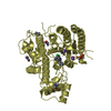
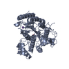
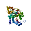
| Title | Crystal structure of Schistosoma mansoni HDAC8 complexed with a triazole hydroxamate inhibitor 2 | |||||||||
 Components Components | Histone deacetylase | |||||||||
 Keywords Keywords | HYDROLASE / Epigenetics / Histone deacetylase / HDAC8 / Selective inhibitor / Pathogen | |||||||||
| Function / homology |  Function and homology information Function and homology information | |||||||||
| Biological species |  Schistosoma mansoni (invertebrata) Schistosoma mansoni (invertebrata) | |||||||||
| Method | X-RAY DIFFRACTION / SYNCHROTRON / MOLECULAR REPLACEMENT / Resolution: 1.61 Å | |||||||||
 Authors Authors | Shaik, T.B. / Romier, C. | |||||||||
| Funding support |  France, 2items France, 2items
| |||||||||
 Citation Citation | Journal: Chemmedchem / Year: 2020 Title: Structure-Based Design, Synthesis, and Biological Evaluation of Triazole-Based smHDAC8 Inhibitors. Authors: Kalinin, D.V. / Jana, S.K. / Pfafenrot, M. / Chakrabarti, A. / Melesina, J. / Shaik, T.B. / Lancelot, J. / Pierce, R.J. / Sippl, W. / Romier, C. / Jung, M. / Holl, R. | |||||||||
| History |
|
- Structure visualization
| Structure viewer | Molecule: MolmilJmol/JSmol |
|---|
- Downloads & links
-Download
| PDBx/mmCIF format | 6tld.cif.gz | 676.6 KB | Display | PDBx/mmCIF format |
|---|---|---|---|---|
| PDB format | pdb6tld.ent.gz | 558.9 KB | Display | PDB format |
| PDBx/mmJSON format | 6tld.json.gz | Tree view | PDBx/mmJSON format | |
| Others |  Other downloads Other downloads |
-Validation report
| Arichive directory | https://data.pdbj.org/pub/pdb/validation_reports/tl/6tldftp://data.pdbj.org/pub/pdb/validation_reports/tl/6tld | HTTPS FTP |
|---|
-Related structure data
| Related structure data | 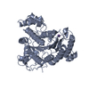 4bz5S S: Starting model for refinement |
|---|---|
| Similar structure data |


-Links
 PDBj
PDBj- Assembly
| Deposited unit | 
| ||||||||
|---|---|---|---|---|---|---|---|---|---|
| 1 | 
| ||||||||
| 2 | 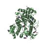
| ||||||||
| 3 | 
| ||||||||
| 4 | 
| ||||||||
| Unit cell |
|
-Components
-Protein , 1 types, 4 molecules ABCD
| #1: Protein | Mass: 50583.027 Da / Num. of mol.: 4 Source method: isolated from a genetically manipulated source Source: (gene. exp.) Schistosoma mansoni (invertebrata) / Gene: HDAC8 / Production host:  Escherichia coli (E. coli) / References: UniProt: A5H660 Escherichia coli (E. coli) / References: UniProt: A5H660 |
|---|
-Non-polymers , 6 types, 1006 molecules 


| #2: Chemical | ChemComp-ZN /  Mass: 65.409 Da / Num. of mol.: 4 / Source method: obtained synthetically / Formula: Zn Mass: 65.409 Da / Num. of mol.: 4 / Source method: obtained synthetically / Formula: Zn#3: Chemical | ChemComp-K /  Mass: 39.098 Da / Num. of mol.: 8 / Source method: obtained synthetically / Formula: K Mass: 39.098 Da / Num. of mol.: 8 / Source method: obtained synthetically / Formula: K#4: Chemical | ChemComp-NK5 / ~{  Mass: 204.185 Da / Num. of mol.: 4 / Source method: obtained synthetically / Formula: C9H8N4O2 / Feature type: SUBJECT OF INVESTIGATION Mass: 204.185 Da / Num. of mol.: 4 / Source method: obtained synthetically / Formula: C9H8N4O2 / Feature type: SUBJECT OF INVESTIGATION#5: Chemical | ChemComp-DMF / Dimethylformamide Mass: 73.094 Da / Num. of mol.: 24 / Source method: obtained synthetically / Formula: C3H7NO Mass: 73.094 Da / Num. of mol.: 24 / Source method: obtained synthetically / Formula: C3H7NO#6: Chemical | ChemComp-GOL / Glycerol Mass: 92.094 Da / Num. of mol.: 6 / Source method: obtained synthetically / Formula: C3H8O3 Mass: 92.094 Da / Num. of mol.: 6 / Source method: obtained synthetically / Formula: C3H8O3#7: Water | ChemComp-HOH / | WaterMass: 18.015 Da / Num. of mol.: 960 / Source method: isolated from a natural source / Formula: H2O |
|---|
-Details
| Has ligand of interest | Y |
|---|
-Experimental details
-Experiment
| Experiment | Method: X-RAY DIFFRACTION / Number of used crystals: 1 |
|---|
- Sample preparation
| Crystal | Density Matthews: 2.29 Å3/Da / Density % sol: 46.39 % |
|---|---|
| Crystal grow | Temperature: 293 K / Method: vapor diffusion, sitting drop / Details: 0.2 M NA,K L-TARTRATE 21% (W/V) PEG3350 |
-Data collection
| Diffraction | Mean temperature: 100 K / Serial crystal experiment: N |
|---|---|
| Diffraction source | Source: SYNCHROTRON / Site: ESRF / Beamline: ID29 / Wavelength: 1.072522 Å |
| Detector | Type: DECTRIS PILATUS 6M / Detector: PIXEL / Date: Feb 24, 2016 |
| Radiation | Protocol: SINGLE WAVELENGTH / Monochromatic (M) / Laue (L): M / Scattering type: x-ray |
| Radiation wavelength | Wavelength: 1.072522 Å / Relative weight: 1 |
| Reflection | Resolution: 1.61→50 Å / Num. obs: 213325 / % possible obs: 92.2 % / Redundancy: 3.6 % / CC1/2: 0.999 / Net I/σ(I): 11.4 |
| Reflection shell | Resolution: 1.61→1.71 Å / Redundancy: 3.5 % / Mean I/σ(I) obs: 1.04 / Num. unique obs: 32281 / CC1/2: 0.498 / % possible all: 86.3 |
- Processing
| Software |
| ||||||||||||||||||||||||||||||||||||||||||||||||||||||||||||||||||||||||||||||||||||||||||||||||||||||||||||||||||||||||||||||||||||||||||||||||||||||||||||||||||||||||||||||||||||||||||
|---|---|---|---|---|---|---|---|---|---|---|---|---|---|---|---|---|---|---|---|---|---|---|---|---|---|---|---|---|---|---|---|---|---|---|---|---|---|---|---|---|---|---|---|---|---|---|---|---|---|---|---|---|---|---|---|---|---|---|---|---|---|---|---|---|---|---|---|---|---|---|---|---|---|---|---|---|---|---|---|---|---|---|---|---|---|---|---|---|---|---|---|---|---|---|---|---|---|---|---|---|---|---|---|---|---|---|---|---|---|---|---|---|---|---|---|---|---|---|---|---|---|---|---|---|---|---|---|---|---|---|---|---|---|---|---|---|---|---|---|---|---|---|---|---|---|---|---|---|---|---|---|---|---|---|---|---|---|---|---|---|---|---|---|---|---|---|---|---|---|---|---|---|---|---|---|---|---|---|---|---|---|---|---|---|---|---|---|
| Refinement | Method to determine structure: MOLECULAR REPLACEMENT Starting model: 4BZ5 Resolution: 1.61→50 Å / SU ML: 0.21 / Cross valid method: THROUGHOUT / σ(F): 1.94 / Phase error: 21.6
| ||||||||||||||||||||||||||||||||||||||||||||||||||||||||||||||||||||||||||||||||||||||||||||||||||||||||||||||||||||||||||||||||||||||||||||||||||||||||||||||||||||||||||||||||||||||||||
| Solvent computation | Shrinkage radii: 0.9 Å / VDW probe radii: 1.11 Å | ||||||||||||||||||||||||||||||||||||||||||||||||||||||||||||||||||||||||||||||||||||||||||||||||||||||||||||||||||||||||||||||||||||||||||||||||||||||||||||||||||||||||||||||||||||||||||
| Displacement parameters | Biso max: 106.63 Å2 / Biso mean: 32.2414 Å2 / Biso min: 14.22 Å2 | ||||||||||||||||||||||||||||||||||||||||||||||||||||||||||||||||||||||||||||||||||||||||||||||||||||||||||||||||||||||||||||||||||||||||||||||||||||||||||||||||||||||||||||||||||||||||||
| Refinement step | Cycle: final / Resolution: 1.61→50 Å
| ||||||||||||||||||||||||||||||||||||||||||||||||||||||||||||||||||||||||||||||||||||||||||||||||||||||||||||||||||||||||||||||||||||||||||||||||||||||||||||||||||||||||||||||||||||||||||
| Refine LS restraints |
| ||||||||||||||||||||||||||||||||||||||||||||||||||||||||||||||||||||||||||||||||||||||||||||||||||||||||||||||||||||||||||||||||||||||||||||||||||||||||||||||||||||||||||||||||||||||||||
| LS refinement shell | Refine-ID: X-RAY DIFFRACTION / Rfactor Rfree error: 0
| ||||||||||||||||||||||||||||||||||||||||||||||||||||||||||||||||||||||||||||||||||||||||||||||||||||||||||||||||||||||||||||||||||||||||||||||||||||||||||||||||||||||||||||||||||||||||||
| Refinement TLS params. | Method: refined / Refine-ID: X-RAY DIFFRACTION
| ||||||||||||||||||||||||||||||||||||||||||||||||||||||||||||||||||||||||||||||||||||||||||||||||||||||||||||||||||||||||||||||||||||||||||||||||||||||||||||||||||||||||||||||||||||||||||
| Refinement TLS group |
|
