 Movie
Movie Controller

[English] 日本語
 Yorodumi
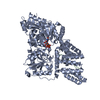
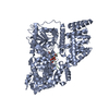
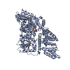
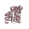
- PDB-6tka: Crystal structure of human O-GlcNAc transferase bound to substrat... -
+ Open data

- Basic information
| Entry | Database: PDB / ID: 6tka | ||||||
|---|---|---|---|---|---|---|---|
| Title | Crystal structure of human O-GlcNAc transferase bound to substrate 7 and a peptide from HCF-1 pro-repeat 2 (11-26) | ||||||
 Components Components |
| ||||||
 Keywords Keywords |  TRANSFERASE / Transferase Complex O-GlcNAc OGT TRANSFERASE / Transferase Complex O-GlcNAc OGT | ||||||
| Function / homology |  Function and homology informationprotein N-acetylglucosaminyltransferase complex / protein O-GlcNAc transferase / regulation of insulin receptor signaling pathway / protein O-acetylglucosaminyltransferase activity / positive regulation of transcription from RNA polymerase II promoter by glucose / acetylglucosaminyltransferase activity / regulation of necroptotic process / regulation of Rac protein signal transduction / negative regulation of stem cell population maintenance / protein O-linked glycosylation ...protein N-acetylglucosaminyltransferase complex / protein O-GlcNAc transferase / regulation of insulin receptor signaling pathway / protein O-acetylglucosaminyltransferase activity / positive regulation of transcription from RNA polymerase II promoter by glucose / acetylglucosaminyltransferase activity / regulation of necroptotic process / regulation of Rac protein signal transduction / negative regulation of stem cell population maintenance / protein O-linked glycosylation / NSL complex / regulation of glycolytic process / regulation of neurotransmitter receptor localization to postsynaptic specialization membrane / RIPK1-mediated regulated necrosis / regulation of synapse assembly / regulation of gluconeogenesis / positive regulation of stem cell population maintenance / Formation of WDR5-containing histone-modifying complexes / positive regulation of proteolysis / phosphatidylinositol-3,4,5-trisphosphate binding / mitophagy / hemopoiesis / negative regulation of proteasomal ubiquitin-dependent protein catabolic process / histone acetyltransferase complex / positive regulation of lipid biosynthetic process / negative regulation of protein ubiquitination / positive regulation of TORC1 signaling / negative regulation of cell migration / response to nutrient / cell projection / positive regulation of translation / mitochondrial membrane / cellular response to glucose stimulus / negative regulation of transforming growth factor beta receptor signaling pathway / circadian regulation of gene expression / response to insulin / Regulation of necroptotic cell death / protein processing / chromatin DNA binding / UCH proteinases / positive regulation of cold-induced thermogenesis / chromatin organization / HATs acetylate histones / glutamatergic synapse / apoptotic process / regulation of transcription by RNA polymerase II / positive regulation of DNA-templated transcription / negative regulation of transcription by RNA polymerase II / signal transduction / positive regulation of transcription by RNA polymerase II / protein-containing complex / nucleoplasm / nucleus / plasma membrane / cytosol Function and homology informationprotein N-acetylglucosaminyltransferase complex / protein O-GlcNAc transferase / regulation of insulin receptor signaling pathway / protein O-acetylglucosaminyltransferase activity / positive regulation of transcription from RNA polymerase II promoter by glucose / acetylglucosaminyltransferase activity / regulation of necroptotic process / regulation of Rac protein signal transduction / negative regulation of stem cell population maintenance / protein O-linked glycosylation ...protein N-acetylglucosaminyltransferase complex / protein O-GlcNAc transferase / regulation of insulin receptor signaling pathway / protein O-acetylglucosaminyltransferase activity / positive regulation of transcription from RNA polymerase II promoter by glucose / acetylglucosaminyltransferase activity / regulation of necroptotic process / regulation of Rac protein signal transduction / negative regulation of stem cell population maintenance / protein O-linked glycosylation / NSL complex / regulation of glycolytic process / regulation of neurotransmitter receptor localization to postsynaptic specialization membrane / RIPK1-mediated regulated necrosis / regulation of synapse assembly / regulation of gluconeogenesis / positive regulation of stem cell population maintenance / Formation of WDR5-containing histone-modifying complexes / positive regulation of proteolysis / phosphatidylinositol-3,4,5-trisphosphate binding / mitophagy / hemopoiesis / negative regulation of proteasomal ubiquitin-dependent protein catabolic process / histone acetyltransferase complex / positive regulation of lipid biosynthetic process / negative regulation of protein ubiquitination / positive regulation of TORC1 signaling / negative regulation of cell migration / response to nutrient / cell projection / positive regulation of translation / mitochondrial membrane / cellular response to glucose stimulus / negative regulation of transforming growth factor beta receptor signaling pathway / circadian regulation of gene expression / response to insulin / Regulation of necroptotic cell death / protein processing / chromatin DNA binding / UCH proteinases / positive regulation of cold-induced thermogenesis / chromatin organization / HATs acetylate histones / glutamatergic synapse / apoptotic process / regulation of transcription by RNA polymerase II / positive regulation of DNA-templated transcription / negative regulation of transcription by RNA polymerase II / signal transduction / positive regulation of transcription by RNA polymerase II / protein-containing complex / nucleoplasm / nucleus / plasma membrane / cytosolSimilarity search - Function | ||||||
| Biological species |  Homo sapiens (human) Homo sapiens (human) | ||||||
| Method | X-RAY DIFFRACTION / SYNCHROTRON / MOLECULAR REPLACEMENT / Resolution: 1.91 Å | ||||||
 Authors Authors | Meek, R.W. / Davies, G.J. | ||||||
| Funding support |  United Kingdom, 1items United Kingdom, 1items
| ||||||
 Citation Citation | Journal: Angew.Chem.Int.Ed.Engl. / Year: 2020 Title: A Direct Fluorescent Activity Assay for Glycosyltransferases Enables Convenient High-Throughput Screening: Application to O-GlcNAc Transferase. Authors: Alteen, M.G. / Gros, C. / Meek, R.W. / Cardoso, D.A. / Busmann, J.A. / Sangouard, G. / Deen, M.C. / Tan, H.Y. / Shen, D.L. / Russell, C.C. / Davies, G.J. / Robinson, P.J. / McCluskey, A. / Vocadlo, D.J. | ||||||
| History |
|
- Structure visualization
| Structure viewer | Molecule: MolmilJmol/JSmol |
|---|
- Downloads & links
-Download
| PDBx/mmCIF format | 6tka.cif.gz | 276.9 KB | Display | PDBx/mmCIF format |
|---|---|---|---|---|
| PDB format | pdb6tka.ent.gz | Display | PDB format | |
| PDBx/mmJSON format | 6tka.json.gz | Tree view | PDBx/mmJSON format | |
| Others |  Other downloads Other downloads |
-Validation report
| Arichive directory | https://data.pdbj.org/pub/pdb/validation_reports/tk/6tkaftp://data.pdbj.org/pub/pdb/validation_reports/tk/6tka | HTTPS FTP |
|---|
-Related structure data
| Related structure data |  4n39S S: Starting model for refinement |
|---|---|
| Similar structure data |


-Links
 PDBj
PDBj


- Assembly
| Deposited unit | 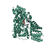
| ||||||||
|---|---|---|---|---|---|---|---|---|---|
| 1 |
| ||||||||
| 2 | 
| ||||||||
| Unit cell |
| ||||||||
| Components on special symmetry positions |
|
-Components
| #1: Protein | Mass: 81388.969 Da / Num. of mol.: 1 Source method: isolated from a genetically manipulated source Source: (gene. exp.) Homo sapiens (human) / Gene: OGT / Production host:  Escherichia coli BL21(DE3) (bacteria) / References: UniProt: O15294, protein O-GlcNAc transferase Escherichia coli BL21(DE3) (bacteria) / References: UniProt: O15294, protein O-GlcNAc transferase |
|---|---|
| #2: Protein/peptide | Mass: 1608.597 Da / Num. of mol.: 1 / Source method: obtained synthetically / Source: (synth.) Homo sapiens (human) |
| #3: Chemical | ChemComp-NJ5 / [[(  Mass: 748.565 Da / Num. of mol.: 1 / Source method: obtained synthetically / Formula: C25H42N4O18P2 / Feature type: SUBJECT OF INVESTIGATION Mass: 748.565 Da / Num. of mol.: 1 / Source method: obtained synthetically / Formula: C25H42N4O18P2 / Feature type: SUBJECT OF INVESTIGATION |
| #4: Chemical | ChemComp-NG8 /   Mass: 320.165 Da / Num. of mol.: 1 / Source method: obtained synthetically / Formula: C16H21BF2N3O Mass: 320.165 Da / Num. of mol.: 1 / Source method: obtained synthetically / Formula: C16H21BF2N3O |
| #5: Water | ChemComp-HOH / Water Mass: 18.015 Da / Num. of mol.: 208 / Source method: isolated from a natural source / Formula: H2O Mass: 18.015 Da / Num. of mol.: 208 / Source method: isolated from a natural source / Formula: H2O |
| Has ligand of interest | Y |
-Experimental details
-Experiment
| Experiment | Method: X-RAY DIFFRACTION / Number of used crystals: 1 |
|---|
- Sample preparation
| Crystal | Density Matthews: 2.37 Å3/Da / Density % sol: 48.06 % |
|---|---|
| Crystal grow | Temperature: 293 K / Method: vapor diffusion, sitting drop / Details: 1.45 M Potassium Sodium Tartrate 0.1 M Tris pH 8.5 |
-Data collection
| Diffraction | Mean temperature: 100 K / Serial crystal experiment: N |
|---|---|
| Diffraction source | Source: SYNCHROTRON / Site: Diamond / Beamline: I04-1 / Wavelength: 0.9159 Å |
| Detector | Type: DECTRIS PILATUS 6M-F / Detector: PIXEL / Date: May 4, 2019 |
| Radiation | Protocol: SINGLE WAVELENGTH / Monochromatic (M) / Laue (L): M / Scattering type: x-ray |
| Radiation wavelength | Wavelength: 0.9159 Å / Relative weight: 1 |
| Reflection | Resolution: 1.91→87.32 Å / Num. obs: 61015 / % possible obs: 100 % / Redundancy: 12 % / CC1/2: 1 / Rmerge(I) obs: 0.1 / Rpim(I) all: 0.03 / Rrim(I) all: 0.104 / Χ2: 0.95 / Net I/σ(I): 15.6 |
| Reflection shell | Resolution: 1.91→1.96 Å / Redundancy: 12.3 % / Mean I/σ(I) obs: 1.1 / Num. unique obs: 4473 / CC1/2: 0.405 / Rpim(I) all: 0.71 / % possible all: 100 |
- Processing
| Software |
| |||||||||||||||||||||||||||||||||||||||||||||||||||||||||||||||||||||||||||||||||||||||||||||||||||||||||||||||||||||||||||||||||||||||||||||||||||||||||||
|---|---|---|---|---|---|---|---|---|---|---|---|---|---|---|---|---|---|---|---|---|---|---|---|---|---|---|---|---|---|---|---|---|---|---|---|---|---|---|---|---|---|---|---|---|---|---|---|---|---|---|---|---|---|---|---|---|---|---|---|---|---|---|---|---|---|---|---|---|---|---|---|---|---|---|---|---|---|---|---|---|---|---|---|---|---|---|---|---|---|---|---|---|---|---|---|---|---|---|---|---|---|---|---|---|---|---|---|---|---|---|---|---|---|---|---|---|---|---|---|---|---|---|---|---|---|---|---|---|---|---|---|---|---|---|---|---|---|---|---|---|---|---|---|---|---|---|---|---|---|---|---|---|---|---|---|---|
| Refinement | Method to determine structure: MOLECULAR REPLACEMENT Starting model: 4n39 Resolution: 1.91→72.95 Å / Cor.coef. Fo:Fc: 0.969 / Cor.coef. Fo:Fc free: 0.951 / Cross valid method: FREE R-VALUE / ESU R: 0.149 / ESU R Free: 0.145 Details: Hydrogens have been added in their riding positions
| |||||||||||||||||||||||||||||||||||||||||||||||||||||||||||||||||||||||||||||||||||||||||||||||||||||||||||||||||||||||||||||||||||||||||||||||||||||||||||
| Solvent computation | Ion probe radii: 0.8 Å / Shrinkage radii: 0.8 Å / VDW probe radii: 1.2 Å | |||||||||||||||||||||||||||||||||||||||||||||||||||||||||||||||||||||||||||||||||||||||||||||||||||||||||||||||||||||||||||||||||||||||||||||||||||||||||||
| Displacement parameters | Biso mean: 38.52 Å2
| |||||||||||||||||||||||||||||||||||||||||||||||||||||||||||||||||||||||||||||||||||||||||||||||||||||||||||||||||||||||||||||||||||||||||||||||||||||||||||
| Refinement step | Cycle: LAST / Resolution: 1.91→72.95 Å
| |||||||||||||||||||||||||||||||||||||||||||||||||||||||||||||||||||||||||||||||||||||||||||||||||||||||||||||||||||||||||||||||||||||||||||||||||||||||||||
| Refine LS restraints |
| |||||||||||||||||||||||||||||||||||||||||||||||||||||||||||||||||||||||||||||||||||||||||||||||||||||||||||||||||||||||||||||||||||||||||||||||||||||||||||
| LS refinement shell |
|
