 Movie
Movie Controller


+ Open data

- Basic information
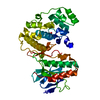
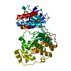
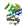
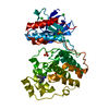
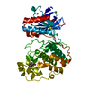
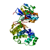
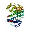
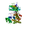
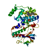
| Entry | Database: PDB / ID: 6sot | ||||||
|---|---|---|---|---|---|---|---|
| Title | Fragment N11290a in complex with MAP kinase p38-alpha | ||||||
 Components Components | Mitogen-activated protein kinase 14 MAPK14 MAPK14 | ||||||
 Keywords Keywords | TRANSFERASE / FBDD / Fragment Based Drug Design / P38 / MAPK14 / Kinase / Transferase. | ||||||
| Function / homology |  Function and homology information Function and homology informationDSCAM interactions / p38MAPK events / Activation of the AP-1 family of transcription factors / Platelet sensitization by LDL / RHO GTPases Activate NADPH Oxidases / ERK/MAPK targets / activated TAK1 mediates p38 MAPK activation / NOD1/2 Signaling Pathway / ADP signalling through P2Y purinoceptor 1 / Oxidative Stress Induced Senescence ...DSCAM interactions / p38MAPK events / Activation of the AP-1 family of transcription factors / Platelet sensitization by LDL / RHO GTPases Activate NADPH Oxidases / ERK/MAPK targets / activated TAK1 mediates p38 MAPK activation / NOD1/2 Signaling Pathway / ADP signalling through P2Y purinoceptor 1 / Oxidative Stress Induced Senescence / Regulation of TP53 Activity through Phosphorylation / Myogenesis / VEGFA-VEGFR2 Pathway / regulation of synaptic membrane adhesion / stress-induced premature senescence / cell surface receptor protein serine/threonine kinase signaling pathway / cartilage condensation / cellular response to UV-B / stress-activated protein kinase signaling cascade / mitogen-activated protein kinase p38 binding / positive regulation of myoblast fusion / negative regulation of hippo signaling / positive regulation of myotube differentiation / NFAT protein binding / glucose import / regulation of cytokine production involved in inflammatory response / p38MAPK cascade / fatty acid oxidation / cellular response to lipoteichoic acid / response to dietary excess / response to muramyl dipeptide / regulation of ossification / MAP kinase activity / mitogen-activated protein kinase / cellular response to vascular endothelial growth factor stimulus / positive regulation of myoblast differentiation / chondrocyte differentiation / vascular endothelial growth factor receptor signaling pathway / stress-activated MAPK cascade / signal transduction in response to DNA damage / skeletal muscle tissue development / lipopolysaccharide-mediated signaling pathway / positive regulation of cardiac muscle cell proliferation / striated muscle cell differentiation / response to muscle stretch / Neutrophil degranulation / positive regulation of brown fat cell differentiation / positive regulation of interleukin-12 production / osteoclast differentiation / positive regulation of erythrocyte differentiation / placenta development / DNA damage checkpoint signaling / cellular response to ionizing radiation / stem cell differentiation / positive regulation of glucose import / response to insulin / bone development / cell morphogenesis / negative regulation of canonical Wnt signaling pathway / cellular response to virus / osteoblast differentiation / spindle pole / positive regulation of protein import into nucleus / glucose metabolic process / positive regulation of reactive oxygen species metabolic process / MAPK cascade / cellular response to tumor necrosis factor / kinase activity / peptidyl-serine phosphorylation / protein phosphatase binding / angiogenesis / cellular response to lipopolysaccharide / response to lipopolysaccharide / transcription by RNA polymerase II / protein kinase activity / intracellular signal transduction / nuclear speck / protein serine kinase activity / protein serine/threonine kinase activity / glutamatergic synapse / apoptotic process / DNA damage response / positive regulation of gene expression / regulation of DNA-templated transcription / regulation of transcription by RNA polymerase II / enzyme binding / positive regulation of transcription by RNA polymerase II / mitochondrion / nucleoplasm / ATP binding / nucleus / cytosol / cytoplasmSimilarity search - Function | ||||||
| Biological species |  Mus musculus (house mouse) Mus musculus (house mouse) | ||||||
| Method | X-RAY DIFFRACTION / SYNCHROTRON / MOLECULAR REPLACEMENT / Resolution: 1.54 Å | ||||||
 Authors Authors | Nichols, C.E. / De Nicola, G.F. | ||||||
| Funding support |  United Kingdom, 1items United Kingdom, 1items
| ||||||
 Citation Citation | Journal: J.Med.Chem. / Year: 2020 Title: Mining the PDB for Tractable Cases Where X-ray Crystallography Combined with Fragment Screens Can Be Used to Systematically Design Protein-Protein Inhibitors: Two Test Cases Illustrated by IL1 ...Title: Mining the PDB for Tractable Cases Where X-ray Crystallography Combined with Fragment Screens Can Be Used to Systematically Design Protein-Protein Inhibitors: Two Test Cases Illustrated by IL1 beta-IL1R and p38 alpha-TAB1 Complexes. Authors: Nichols, C. / Ng, J. / Keshu, A. / Kelly, G. / Conte, M.R. / Marber, M.S. / Fraternali, F. / De Nicola, G.F. | ||||||
| History |
|
- Structure visualization
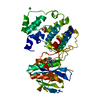
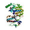
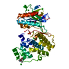
| Structure viewer | Molecule: MolmilJmol/JSmol |
|---|
- Downloads & links
-Download
| PDBx/mmCIF format | 6sot.cif.gz | 111.3 KB | Display | PDBx/mmCIF format |
|---|---|---|---|---|
| PDB format | pdb6sot.ent.gz | 67.2 KB | Display | PDB format |
| PDBx/mmJSON format | 6sot.json.gz | Tree view | PDBx/mmJSON format | |
| Others |  Other downloads Other downloads |
-Validation report
| Arichive directory | https://data.pdbj.org/pub/pdb/validation_reports/so/6sotftp://data.pdbj.org/pub/pdb/validation_reports/so/6sot | HTTPS FTP |
|---|
-Related structure data
| Related structure data |  5r85C  5r86C 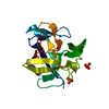 5r87C 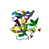 5r88C  5r89C  5r8aC  5r8bC 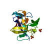 5r8cC 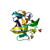 5r8dC 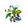 5r8eC 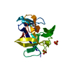 5r8fC  5r8gC  5r8hC  5r8iC 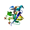 5r8jC 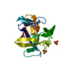 5r8kC  5r8lC  5r8mC  5r8nC 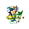 5r8oC 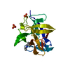 5r8pC 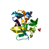 5r8qC 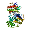 5r8uC 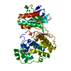 5r8vC  5r8wC  5r8xC 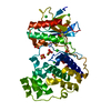 5r8yC  5r8zC 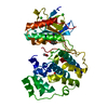 5r90C  5r91C  5r92C 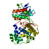 5r93C 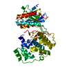 5r94C 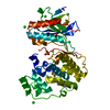 5r95C 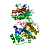 5r96C 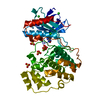 5r97C 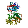 5r98C 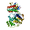 5r99C 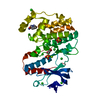 5r9aC 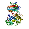 5r9bC 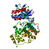 5r9cC  5r9dC 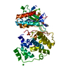 5r9eC 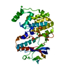 5r9fC  5r9gC  5r9hC 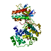 5r9iC 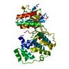 5r9jC  5r9kC  5r9lC 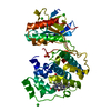 5r9mC 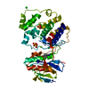 5r9nC 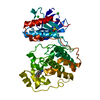 5r9oC  5r9pC 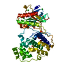 5r9qC 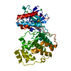 5r9rC 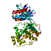 5r9sC 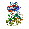 5r9tC 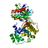 5r9uC  5r9vC 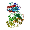 5r9wC 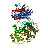 5r9xC 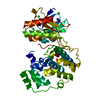 5r9yC  5r9zC 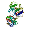 5ra0C 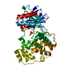 5ra1C 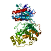 5ra2C 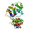 5ra3C  5ra4C 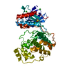 5ra5C 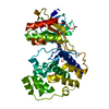 5ra6C 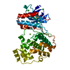 5ra7C 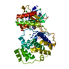 5ra8C 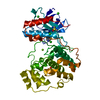 5ra9C  6so1SC 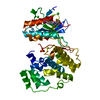 6so2C  6so4C 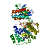 6sodC 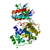 6soiC 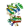 6souC  6sovC 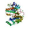 6sp9C 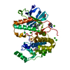 6splC 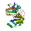 6y7wC 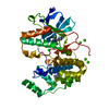 6y7xC 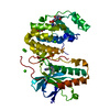 6y7yC  6y80C 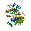 6y81C 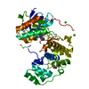 6ycuC 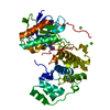 6ycwC S: Starting model for refinement C: citing same article ( |
|---|---|
| Similar structure data |


-Links
 PDBj
PDBj


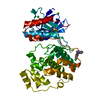
- Assembly
| Deposited unit | 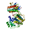
| ||||||||||||
|---|---|---|---|---|---|---|---|---|---|---|---|---|---|
| 1 |
| ||||||||||||
| Unit cell |
|
-Components
-Protein , 1 types, 1 molecules A
| #1: Protein | MAPK14 / MAPK 14 / CRK1 / Mitogen-activated protein kinase p38 alpha / MAP kinase p38 alpha Mass: 43808.738 Da / Num. of mol.: 1 Source method: isolated from a genetically manipulated source Source: (gene. exp.) Mus musculus (house mouse) / Gene: Mapk14, Crk1, Csbp1, Csbp2 / Production host:  Escherichia coli (E. coli) Escherichia coli (E. coli)References: UniProt: P47811, mitogen-activated protein kinase |
|---|
-Non-polymers , 6 types, 286 molecules 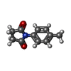


| #2: Chemical | ChemComp-LOQ /  Mass: 189.211 Da / Num. of mol.: 1 / Source method: obtained synthetically / Formula: C11H11NO2 / Feature type: SUBJECT OF INVESTIGATION Mass: 189.211 Da / Num. of mol.: 1 / Source method: obtained synthetically / Formula: C11H11NO2 / Feature type: SUBJECT OF INVESTIGATION | ||||||||
|---|---|---|---|---|---|---|---|---|---|
| #3: Chemical | ChemComp-CL / Chloride Mass: 35.453 Da / Num. of mol.: 5 / Source method: obtained synthetically / Formula: Cl Mass: 35.453 Da / Num. of mol.: 5 / Source method: obtained synthetically / Formula: Cl#4: Chemical | ChemComp-MG / |  Mass: 24.305 Da / Num. of mol.: 1 / Source method: obtained synthetically / Formula: Mg Mass: 24.305 Da / Num. of mol.: 1 / Source method: obtained synthetically / Formula: Mg#5: Chemical | Sulfate Mass: 96.063 Da / Num. of mol.: 2 / Source method: obtained synthetically / Formula: SO4 Mass: 96.063 Da / Num. of mol.: 2 / Source method: obtained synthetically / Formula: SO4#6: Chemical | Ethylene glycol Mass: 62.068 Da / Num. of mol.: 2 / Source method: obtained synthetically / Formula: C2H6O2 Mass: 62.068 Da / Num. of mol.: 2 / Source method: obtained synthetically / Formula: C2H6O2#7: Water | ChemComp-HOH / | WaterMass: 18.015 Da / Num. of mol.: 275 / Source method: isolated from a natural source / Formula: H2O |
-Details
| Has ligand of interest | Y |
|---|
-Experimental details
-Experiment
| Experiment | Method: X-RAY DIFFRACTION / Number of used crystals: 1 |
|---|
- Sample preparation
| Crystal | Density Matthews: 3.19 Å3/Da / Density % sol: 61.49 % |
|---|---|
| Crystal grow | Temperature: 291 K / Method: vapor diffusion, sitting drop Details: 27.5% PEG3350, 0.1M magnesium chloride, 0.1M magnesium sulphate, 0.1M bis-tris propane, 10% glycerol |
-Data collection
| Diffraction | Mean temperature: 100 K / Serial crystal experiment: N |
|---|---|
| Diffraction source | Source: SYNCHROTRON / Site: Diamond / Beamline: I04-1 / Wavelength: 0.92 Å |
| Detector | Type: DECTRIS PILATUS 6M-F / Detector: PIXEL / Date: Feb 12, 2016 |
| Radiation | Protocol: SINGLE WAVELENGTH / Monochromatic (M) / Laue (L): M / Scattering type: x-ray |
| Radiation wavelength | Wavelength: 0.92 Å / Relative weight: 1 |
| Reflection | Resolution: 1.54→85.81 Å / Num. obs: 75111 / % possible obs: 99.7 % / Redundancy: 6.3 % / Biso Wilson estimate: 19.12 Å2 / CC1/2: 0.999 / Net I/σ(I): 11.3 |
| Reflection shell | Resolution: 1.54→1.58 Å / Redundancy: 5.4 % / Mean I/σ(I) obs: 1 / Num. unique obs: 5444 / CC1/2: 0.525 / % possible all: 99.5 |
- Processing
| Software |
| |||||||||||||||||||||||||||||||||||||||||||||||||||||||||||||||||||||||||||||||||||||||||||||||||||||||||||||||||||||||||||||||||||||||||||||||||||||||||||||||||||||||||||||||||||||||||||||||||||||||||||||||||||||||||
|---|---|---|---|---|---|---|---|---|---|---|---|---|---|---|---|---|---|---|---|---|---|---|---|---|---|---|---|---|---|---|---|---|---|---|---|---|---|---|---|---|---|---|---|---|---|---|---|---|---|---|---|---|---|---|---|---|---|---|---|---|---|---|---|---|---|---|---|---|---|---|---|---|---|---|---|---|---|---|---|---|---|---|---|---|---|---|---|---|---|---|---|---|---|---|---|---|---|---|---|---|---|---|---|---|---|---|---|---|---|---|---|---|---|---|---|---|---|---|---|---|---|---|---|---|---|---|---|---|---|---|---|---|---|---|---|---|---|---|---|---|---|---|---|---|---|---|---|---|---|---|---|---|---|---|---|---|---|---|---|---|---|---|---|---|---|---|---|---|---|---|---|---|---|---|---|---|---|---|---|---|---|---|---|---|---|---|---|---|---|---|---|---|---|---|---|---|---|---|---|---|---|---|---|---|---|---|---|---|---|---|---|---|---|---|---|---|---|---|
| Refinement | Method to determine structure: MOLECULAR REPLACEMENT Starting model: 6SO1 Resolution: 1.54→71.14 Å / SU ML: 0.2412 / Cross valid method: FREE R-VALUE / σ(F): 0 / Phase error: 26.1826
| |||||||||||||||||||||||||||||||||||||||||||||||||||||||||||||||||||||||||||||||||||||||||||||||||||||||||||||||||||||||||||||||||||||||||||||||||||||||||||||||||||||||||||||||||||||||||||||||||||||||||||||||||||||||||
| Solvent computation | Shrinkage radii: 0.9 Å / VDW probe radii: 1.11 Å | |||||||||||||||||||||||||||||||||||||||||||||||||||||||||||||||||||||||||||||||||||||||||||||||||||||||||||||||||||||||||||||||||||||||||||||||||||||||||||||||||||||||||||||||||||||||||||||||||||||||||||||||||||||||||
| Displacement parameters | Biso mean: 25.11 Å2 | |||||||||||||||||||||||||||||||||||||||||||||||||||||||||||||||||||||||||||||||||||||||||||||||||||||||||||||||||||||||||||||||||||||||||||||||||||||||||||||||||||||||||||||||||||||||||||||||||||||||||||||||||||||||||
| Refinement step | Cycle: LAST / Resolution: 1.54→71.14 Å
| |||||||||||||||||||||||||||||||||||||||||||||||||||||||||||||||||||||||||||||||||||||||||||||||||||||||||||||||||||||||||||||||||||||||||||||||||||||||||||||||||||||||||||||||||||||||||||||||||||||||||||||||||||||||||
| Refine LS restraints |
| |||||||||||||||||||||||||||||||||||||||||||||||||||||||||||||||||||||||||||||||||||||||||||||||||||||||||||||||||||||||||||||||||||||||||||||||||||||||||||||||||||||||||||||||||||||||||||||||||||||||||||||||||||||||||
| LS refinement shell |
|
