 Movie
Movie Controller


+ Open data

- Basic information
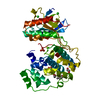
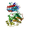
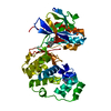
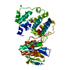
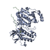
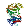
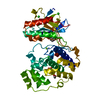
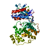
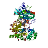
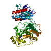
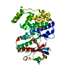
| Entry | Database: PDB / ID: 1r39 | ||||||
|---|---|---|---|---|---|---|---|
| Title | THE STRUCTURE OF P38ALPHA | ||||||
 Components Components | Mitogen-activated protein kinase 14 MAPK14 MAPK14 | ||||||
 Keywords Keywords | TRANSFERASE / SERINE/THREONINE KINASE / MUTAGENESIS / STABILIZATION | ||||||
| Function / homology |  Function and homology information Function and homology informationpositive regulation of cyclase activity / Activation of PPARGC1A (PGC-1alpha) by phosphorylation / CD163 mediating an anti-inflammatory response / regulation of synaptic membrane adhesion / stress-induced premature senescence / cell surface receptor protein serine/threonine kinase signaling pathway / 3'-UTR-mediated mRNA stabilization / KSRP (KHSRP) binds and destabilizes mRNA / DSCAM interactions / cartilage condensation ...positive regulation of cyclase activity / Activation of PPARGC1A (PGC-1alpha) by phosphorylation / CD163 mediating an anti-inflammatory response / regulation of synaptic membrane adhesion / stress-induced premature senescence / cell surface receptor protein serine/threonine kinase signaling pathway / 3'-UTR-mediated mRNA stabilization / KSRP (KHSRP) binds and destabilizes mRNA / DSCAM interactions / cartilage condensation / cellular response to UV-B / Platelet sensitization by LDL / stress-activated protein kinase signaling cascade / mitogen-activated protein kinase p38 binding / positive regulation of myoblast fusion / positive regulation of muscle cell differentiation / negative regulation of hippo signaling / positive regulation of myotube differentiation / NFAT protein binding / Myogenesis / glucose import / Activation of the AP-1 family of transcription factors / ERK/MAPK targets / regulation of cytokine production involved in inflammatory response / p38MAPK cascade / fatty acid oxidation / cellular response to lipoteichoic acid / MAP kinase kinase activity / response to dietary excess / response to muramyl dipeptide / RHO GTPases Activate NADPH Oxidases / regulation of ossification / MAP kinase activity / mitogen-activated protein kinase / cellular response to vascular endothelial growth factor stimulus / positive regulation of myoblast differentiation / chondrocyte differentiation / vascular endothelial growth factor receptor signaling pathway / stress-activated MAPK cascade / signal transduction in response to DNA damage / skeletal muscle tissue development / lipopolysaccharide-mediated signaling pathway / positive regulation of cardiac muscle cell proliferation / negative regulation of inflammatory response to antigenic stimulus / p38MAPK events / striated muscle cell differentiation / response to muscle stretch / positive regulation of brown fat cell differentiation / positive regulation of interleukin-12 production / osteoclast differentiation / positive regulation of erythrocyte differentiation / placenta development / DNA damage checkpoint signaling / activated TAK1 mediates p38 MAPK activation / cellular response to ionizing radiation / stem cell differentiation / positive regulation of glucose import / response to insulin / NOD1/2 Signaling Pathway / bone development / cell morphogenesis / negative regulation of canonical Wnt signaling pathway / platelet activation / cellular response to virus / osteoblast differentiation / VEGFA-VEGFR2 Pathway / spindle pole / positive regulation of protein import into nucleus / ADP signalling through P2Y purinoceptor 1 / glucose metabolic process / positive regulation of reactive oxygen species metabolic process / chemotaxis / cellular senescence / cellular response to tumor necrosis factor / peptidyl-serine phosphorylation / protein phosphatase binding / angiogenesis / secretory granule lumen / Oxidative Stress Induced Senescence / cellular response to lipopolysaccharide / Regulation of TP53 Activity through Phosphorylation / ficolin-1-rich granule lumen / transcription by RNA polymerase II / cell surface receptor signaling pathway / intracellular signal transduction / nuclear speck / protein serine kinase activity / protein serine/threonine kinase activity / glutamatergic synapse / apoptotic process / Neutrophil degranulation / positive regulation of gene expression / regulation of transcription by RNA polymerase II / enzyme binding / signal transduction / positive regulation of transcription by RNA polymerase II / mitochondrion / extracellular region / nucleoplasm / ATP bindingSimilarity search - Function | ||||||
| Biological species |  Homo sapiens (human) Homo sapiens (human) | ||||||
| Method | X-RAY DIFFRACTION / SYNCHROTRON / FOURIER SYNTHESIS / Resolution: 2.3 Å | ||||||
 Authors Authors | Patel, S.B. / Cameron, P.M. / Frantz-Wattley, B. / O'Neill, E. / Becker, J.W. / Scapin, G. | ||||||
 Citation Citation | Journal: Biochim.Biophys.Acta / Year: 2004 Title: Lattice stabilization and enhanced diffraction in human p38 alpha crystals by protein engineering. Authors: Patel, S.B. / Cameron, P.M. / Frantz-Wattley, B. / O'Neill, E. / Becker, J.W. / Scapin, G. | ||||||
| History |
|
- Structure visualization
| Structure viewer | Molecule: MolmilJmol/JSmol |
|---|
- Downloads & links
-Download
| PDBx/mmCIF format | 1r39.cif.gz | 84 KB | Display | PDBx/mmCIF format |
|---|---|---|---|---|
| PDB format | pdb1r39.ent.gz | 62.4 KB | Display | PDB format |
| PDBx/mmJSON format | 1r39.json.gz | Tree view | PDBx/mmJSON format | |
| Others |  Other downloads Other downloads |
-Validation report
| Arichive directory | https://data.pdbj.org/pub/pdb/validation_reports/r3/1r39ftp://data.pdbj.org/pub/pdb/validation_reports/r3/1r39 | HTTPS FTP |
|---|
-Related structure data
| Related structure data | 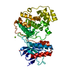 1r3cC 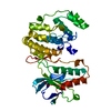 1wfcS C: citing same article ( S: Starting model for refinement |
|---|---|
| Similar structure data |


-Links
 PDBj
PDBj


- Assembly
| Deposited unit | 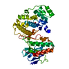
| ||||||||
|---|---|---|---|---|---|---|---|---|---|
| 1 |
| ||||||||
| Unit cell |
|
-Components
| #1: Protein | MAPK14 / Mitogen-activated protein kinase p38alpha / MAP kinase p38alpha / Cytokine suppressive anti- ...Mitogen-activated protein kinase p38alpha / MAP kinase p38alpha / Cytokine suppressive anti-inflammatory drug binding protein / CSAID binding protein / CSBP / MAX-interacting protein 2 / MAP kinase MXI2 / SAPK2A Mass: 41998.938 Da / Num. of mol.: 1 Source method: isolated from a genetically manipulated source Source: (gene. exp.) Homo sapiens (human) / Gene: MAPK14, CSBP1, CSBP2, CSBP OR MXI2 / Plasmid: pET / Species (production host): Escherichia coli / Production host:  Escherichia coli BL21(DE3) (bacteria) / Strain (production host): bl21(DE3) / References: UniProt: Q16539, EC: 2.7.1.37 Escherichia coli BL21(DE3) (bacteria) / Strain (production host): bl21(DE3) / References: UniProt: Q16539, EC: 2.7.1.37 |
|---|---|
| #2: Chemical | ChemComp-SO4 / Sulfate  Mass: 96.063 Da / Num. of mol.: 1 / Source method: obtained synthetically / Formula: SO4 Mass: 96.063 Da / Num. of mol.: 1 / Source method: obtained synthetically / Formula: SO4 |
| #3: Water | ChemComp-HOH / Water Mass: 18.015 Da / Num. of mol.: 127 / Source method: isolated from a natural source / Formula: H2O Mass: 18.015 Da / Num. of mol.: 127 / Source method: isolated from a natural source / Formula: H2O |
-Experimental details
-Experiment
| Experiment | Method: X-RAY DIFFRACTION / Number of used crystals: 1 |
|---|
- Sample preparation
| Crystal | Density Matthews: 2.722 Å3/Da / Density % sol: 52.2 % | |||||||||||||||||||||||||||||||||||||||||||||||||||||||||||||||
|---|---|---|---|---|---|---|---|---|---|---|---|---|---|---|---|---|---|---|---|---|---|---|---|---|---|---|---|---|---|---|---|---|---|---|---|---|---|---|---|---|---|---|---|---|---|---|---|---|---|---|---|---|---|---|---|---|---|---|---|---|---|---|---|---|
| Crystal grow | Temperature: 298 K / Method: vapor diffusion, hanging drop / pH: 7 Details: SODIUM CITRATE, AMMONIUM SULFATE, HEPES, pH 7.00, VAPOR DIFFUSION, HANGING DROP, temperature 298K | |||||||||||||||||||||||||||||||||||||||||||||||||||||||||||||||
| Crystal grow | *PLUS pH: 7.4 / Method: vapor diffusion, hanging drop | |||||||||||||||||||||||||||||||||||||||||||||||||||||||||||||||
| Components of the solutions | *PLUS
|
-Data collection
| Diffraction | Mean temperature: 100 K |
|---|---|
| Diffraction source | Source: SYNCHROTRON / Site: APS  / Beamline: 17-ID / Wavelength: 1 / Wavelength: 1 Å / Beamline: 17-ID / Wavelength: 1 / Wavelength: 1 Å |
| Detector | Type: MARRESEARCH / Detector: CCD / Date: Mar 16, 2000 |
| Radiation | Protocol: SINGLE WAVELENGTH / Monochromatic (M) / Laue (L): M / Scattering type: x-ray |
| Radiation wavelength | Wavelength: 1 Å / Relative weight: 1 |
| Reflection | Resolution: 2.3→15 Å / Num. obs: 19803 / % possible obs: 95.3 % / Observed criterion σ(I): -3 / Redundancy: 8.8 % / Biso Wilson estimate: 41.2 Å2 / Rmerge(I) obs: 0.119 / Net I/σ(I): 6.1 |
| Reflection shell | Resolution: 2.3→2.4 Å / Redundancy: 6.4 % / Rmerge(I) obs: 0.438 / Mean I/σ(I) obs: 1.2 / Num. unique all: 1652 / % possible all: 80.7 |
| Reflection | *PLUS Highest resolution: 2.3 Å / Lowest resolution: 15 Å / Redundancy: 8.8 % |
| Reflection shell | *PLUS Highest resolution: 2.3 Å / Lowest resolution: 2.4 Å / % possible obs: 80.7 % / Redundancy: 6.4 % / Num. unique obs: 1652 / Mean I/σ(I) obs: 1.2 |
- Processing
| Software |
| ||||||||||||||||||||||||||||||||||||||||||||||||||||||||||||||||||||||||||||||||
|---|---|---|---|---|---|---|---|---|---|---|---|---|---|---|---|---|---|---|---|---|---|---|---|---|---|---|---|---|---|---|---|---|---|---|---|---|---|---|---|---|---|---|---|---|---|---|---|---|---|---|---|---|---|---|---|---|---|---|---|---|---|---|---|---|---|---|---|---|---|---|---|---|---|---|---|---|---|---|---|---|---|
| Refinement | Method to determine structure: FOURIER SYNTHESIS Starting model: PDB ENTRY 1WFC Resolution: 2.3→15 Å / Isotropic thermal model: ANISOTROPIC / Cross valid method: THROUGHOUT / σ(F): 0 / Stereochemistry target values: ENGH & HUBER
| ||||||||||||||||||||||||||||||||||||||||||||||||||||||||||||||||||||||||||||||||
| Solvent computation | Solvent model: MASK / Bsol: 41.2 Å2 / ksol: 0.371 e/Å3 | ||||||||||||||||||||||||||||||||||||||||||||||||||||||||||||||||||||||||||||||||
| Displacement parameters | Biso mean: 40.9 Å2
| ||||||||||||||||||||||||||||||||||||||||||||||||||||||||||||||||||||||||||||||||
| Refine analyze |
| ||||||||||||||||||||||||||||||||||||||||||||||||||||||||||||||||||||||||||||||||
| Refinement step | Cycle: LAST / Resolution: 2.3→15 Å
| ||||||||||||||||||||||||||||||||||||||||||||||||||||||||||||||||||||||||||||||||
| Refine LS restraints |
| ||||||||||||||||||||||||||||||||||||||||||||||||||||||||||||||||||||||||||||||||
| LS refinement shell | Resolution: 2.3→2.38 Å
| ||||||||||||||||||||||||||||||||||||||||||||||||||||||||||||||||||||||||||||||||
| Refinement | *PLUS Highest resolution: 2.3 Å / Lowest resolution: 15 Å / % reflection Rfree: 5 % | ||||||||||||||||||||||||||||||||||||||||||||||||||||||||||||||||||||||||||||||||
| Solvent computation | *PLUS | ||||||||||||||||||||||||||||||||||||||||||||||||||||||||||||||||||||||||||||||||
| Displacement parameters | *PLUS | ||||||||||||||||||||||||||||||||||||||||||||||||||||||||||||||||||||||||||||||||
| Refine LS restraints | *PLUS
|
