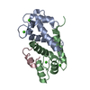
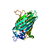
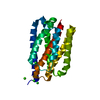
[English] 日本語
Yorodumi
- PDB-6rw2: Bicycle Toxin Conjugate bound to EphA2 -

+
Open data


ID or keywords:

Loading...

-
Basic information

Entry
Database: PDB / ID: 6rw2
TitleBicycle Toxin Conjugate bound to EphA2
Components
  • ALA-ARG-ASP-CYS-PRO-LEU-VAL-ASN-PRO-LEU-CYS-LEU-HIS-PRO-GLY-TRP-THR-CYS
  • Ephrin type-A receptor 2
KeywordsSIGNALING PROTEIN / EphA2 / inhibitor / complex
Function / homology
Function and homology information


notochord cell development / notochord formation / lens fiber cell morphogenesis / blood vessel endothelial cell proliferation involved in sprouting angiogenesis / negative regulation of lymphangiogenesis / axial mesoderm formation / pericyte cell differentiation / cAMP metabolic process / positive regulation of bicellular tight junction assembly / regulation of blood vessel endothelial cell migration ...notochord cell development / notochord formation / lens fiber cell morphogenesis / blood vessel endothelial cell proliferation involved in sprouting angiogenesis / negative regulation of lymphangiogenesis / axial mesoderm formation / pericyte cell differentiation / cAMP metabolic process / positive regulation of bicellular tight junction assembly / regulation of blood vessel endothelial cell migration / leading edge membrane / negative regulation of chemokine production / bone remodeling / transmembrane-ephrin receptor activity / post-anal tail morphogenesis / response to growth factor / activation of GTPase activity / regulation of lamellipodium assembly / tight junction / branching involved in mammary gland duct morphogenesis / EPH-Ephrin signaling / neural tube development / RND1 GTPase cycle / RND2 GTPase cycle / RND3 GTPase cycle / mammary gland epithelial cell proliferation / RHOV GTPase cycle / EPHA-mediated growth cone collapse / growth factor binding / regulation of cell adhesion mediated by integrin / lamellipodium membrane / RHOU GTPase cycle / RHOG GTPase cycle / EPH-ephrin mediated repulsion of cells / RAC2 GTPase cycle / ephrin receptor signaling pathway / RAC3 GTPase cycle / negative regulation of phosphatidylinositol 3-kinase/protein kinase B signal transduction / vasculogenesis / regulation of angiogenesis / keratinocyte differentiation / RAC1 GTPase cycle / transmembrane receptor protein tyrosine kinase activity / cell chemotaxis / negative regulation of angiogenesis / osteoclast differentiation / regulation of ERK1 and ERK2 cascade / phosphatidylinositol 3-kinase/protein kinase B signal transduction / skeletal system development / molecular function activator activity / protein localization to plasma membrane / cell motility / positive regulation of protein localization to plasma membrane / axon guidance / receptor protein-tyrosine kinase / ruffle membrane / osteoblast differentiation / intrinsic apoptotic signaling pathway in response to DNA damage / cell migration / virus receptor activity / lamellipodium / receptor complex / cell adhesion / positive regulation of cell migration / defense response to Gram-positive bacterium / cadherin binding / inflammatory response / phosphorylation / focal adhesion / dendrite / cell surface / ATP binding / plasma membrane
Similarity search - Function
Ephrin type-A receptor 2, ligand binding domain / Ephrin receptor type-A /type-B / Ephrin receptor ligand binding domain / Tyrosine-protein kinase, receptor class V, conserved site / Ephrin receptor, transmembrane domain / Ephrin receptor ligand binding domain / Ephrin type-A receptor 2 transmembrane domain / Receptor tyrosine kinase class V signature 1. / Receptor tyrosine kinase class V signature 2. / Eph receptor ligand-binding domain profile. ...Ephrin type-A receptor 2, ligand binding domain / Ephrin receptor type-A /type-B / Ephrin receptor ligand binding domain / Tyrosine-protein kinase, receptor class V, conserved site / Ephrin receptor, transmembrane domain / Ephrin receptor ligand binding domain / Ephrin type-A receptor 2 transmembrane domain / Receptor tyrosine kinase class V signature 1. / Receptor tyrosine kinase class V signature 2. / Eph receptor ligand-binding domain profile. / Ephrin receptor ligand binding domain / Putative ephrin-receptor like / SAM domain (Sterile alpha motif) / Galactose-binding domain-like / SAM domain profile. / Sterile alpha motif. / Sterile alpha motif domain / Sterile alpha motif/pointed domain superfamily / Growth factor receptor cysteine-rich domain superfamily / Fibronectin type III domain / Fibronectin type 3 domain / Fibronectin type-III domain profile. / Galactose-binding-like domain superfamily / Fibronectin type III / Fibronectin type III superfamily / Tyrosine-protein kinase, catalytic domain / Tyrosine kinase, catalytic domain / Tyrosine protein kinases specific active-site signature. / Tyrosine-protein kinase, active site / Protein tyrosine and serine/threonine kinase / Serine-threonine/tyrosine-protein kinase, catalytic domain / Jelly Rolls / Protein kinase, ATP binding site / Protein kinases ATP-binding region signature. / Immunoglobulin-like fold / Protein kinase domain profile. / Protein kinase domain / Protein kinase-like domain superfamily / Sandwich / Mainly Beta
Similarity search - Domain/homology
Chem-29N / Ephrin type-A receptor 2
Similarity search - Component
Biological speciesHomo sapiens (human)
synthetic construct (others)
MethodX-RAY DIFFRACTION / SYNCHROTRON / MOLECULAR REPLACEMENT / Resolution: 2.26 Å
AuthorsBrown, D.G. / Schroeder, S. / Chen, L.
CitationJournal: J.Med.Chem. / Year: 2020
Title: Identification and Optimization of EphA2-Selective Bicycles for the Delivery of Cytotoxic Payloads.
Authors: Mudd, G.E. / Brown, A. / Chen, L. / van Rietschoten, K. / Watcham, S. / Teufel, D.P. / Pavan, S. / Lani, R. / Huxley, P. / Bennett, G.S.
History
DepositionJun 3, 2019Deposition site: PDBE / Processing site: PDBE
Revision 1.0Apr 8, 2020Provider: repository / Type: Initial release
Revision 1.1Apr 22, 2020Group: Database references / Category: citation / citation_author
Item: _citation.title / _citation_author.identifier_ORCID / _citation_author.name
Revision 1.2May 6, 2020Group: Database references / Category: citation / citation_author
Item: _citation.journal_volume / _citation.page_first ..._citation.journal_volume / _citation.page_first / _citation.page_last / _citation_author.identifier_ORCID

-
Structure visualization

Structure viewerMolecule:
MolmilJmol/JSmol

Downloads & links

-
Assembly

Deposited unit
A: Ephrin type-A receptor 2
B: ALA-ARG-ASP-CYS-PRO-LEU-VAL-ASN-PRO-LEU-CYS-LEU-HIS-PRO-GLY-TRP-THR-CYS
hetero molecules


Theoretical massNumber of molelcules
Total (without water)24,3514
Polymers24,0042
Non-polymers3472
Water77543
1


  • Idetical with deposited unit
  • defined by author&software
  • Evidence: gel filtration
TypeNameSymmetry operationNumber
identity operation1_555x,y,z1
Buried area2050 Å2
ΔGint-15 kcal/mol
Surface area9810 Å2
MethodPISA
Unit cell
Length a, b, c (Å)42.090, 113.680, 130.960
Angle α, β, γ (deg.)90.000, 90.000, 90.000
Int Tables number23
Space group name H-MI222

-
Components

#1: Protein Ephrin type-A receptor 2 / Epithelial cell kinase / Tyrosine-protein kinase receptor ECK


Mass: 22006.697 Da / Num. of mol.: 1
Source method: isolated from a genetically manipulated source
Source: (gene. exp.) Homo sapiens (human) / Gene: EPHA2, ECK / Production host: Escherichia coli (E. coli)
References: UniProt: P29317, receptor protein-tyrosine kinase
#2: Protein/peptide ALA-ARG-ASP-CYS-PRO-LEU-VAL-ASN-PRO-LEU-CYS-LEU-HIS-PRO-GLY-TRP-THR-CYS


Mass: 1997.366 Da / Num. of mol.: 1 / Source method: obtained synthetically / Source: (synth.) synthetic construct (others)
#3: Chemical ChemComp-GOL / GLYCEROL / GLYCERIN / PROPANE-1,2,3-TRIOL / Glycerol


Mass: 92.094 Da / Num. of mol.: 1 / Source method: obtained synthetically / Formula: C3H8O3
#4: Chemical ChemComp-29N / 1,1',1''-(1,3,5-triazinane-1,3,5-triyl)tripropan-1-one / 1,1',1''-(1,3,5-triazinane-1,3,5-triyl)triprop-2-en-1-one, bound form


Mass: 255.313 Da / Num. of mol.: 1 / Source method: obtained synthetically / Formula: C12H21N3O3 / Feature type: SUBJECT OF INVESTIGATION
#5: Water ChemComp-HOH / water / Water


Mass: 18.015 Da / Num. of mol.: 43 / Source method: isolated from a natural source / Formula: H2O

-
Experimental details

-
Experiment

ExperimentMethod: X-RAY DIFFRACTION / Number of used crystals: 1

-
Sample preparation

CrystalDensity Matthews: 3.55 Å3/Da / Density % sol: 65.39 %
Crystal growTemperature: 293 K / Method: vapor diffusion, hanging drop
Details: 0.1M sodium acetate 0.2M lithium sulphate 8-10% PEG 6000
PH range: 5.5-5.75

-
Data collection

DiffractionMean temperature: 100 K / Serial crystal experiment: N
Diffraction sourceSource: SYNCHROTRON / Site: Diamond / Beamline: I04 / Wavelength: 0.9795 Å
DetectorType: DECTRIS PILATUS 6M / Detector: PIXEL / Date: Feb 3, 2017
RadiationProtocol: SINGLE WAVELENGTH / Monochromatic (M) / Laue (L): M / Scattering type: x-ray
Radiation wavelengthWavelength: 0.9795 Å / Relative weight: 1
ReflectionResolution: 2.26→58.64 Å / Num. obs: 15014 / % possible obs: 98.7 % / Redundancy: 5.8 % / Rmerge(I) obs: 0.077 / Net I/σ(I): 15.8
Reflection shellResolution: 2.26→2.33 Å / Redundancy: 3.3 % / Rmerge(I) obs: 0.935 / Num. unique obs: 667 / % possible all: 87.6

-
Processing

Software
NameVersionClassification
REFMAC5.8.0238refinement
PDB_EXTRACT3.22data extraction
xia2data reduction
Aimlessdata scaling
PHASERphasing
RefinementMethod to determine structure: MOLECULAR REPLACEMENT / Resolution: 2.26→56.9 Å / Cor.coef. Fo:Fc: 0.949 / Cor.coef. Fo:Fc free: 0.903 / Cross valid method: THROUGHOUT / σ(F): 0 / ESU R: 0.225 / ESU R Free: 0.207
Details: HYDROGENS HAVE BEEN ADDED IN THE RIDING POSITIONS U VALUES : REFINED INDIVIDUALLY
RfactorNum. reflection% reflectionSelection details
Rfree0.2646 780 5.2 %RANDOM
Rwork0.211 ---
obs0.2138 14234 98.68 %-
Solvent computationIon probe radii: 0.8 Å / Shrinkage radii: 0.8 Å / VDW probe radii: 1.2 Å
Displacement parametersBiso max: 131.32 Å2 / Biso mean: 41.752 Å2 / Biso min: 20.17 Å2
Baniso -1Baniso -2Baniso -3
1--1.2 Å2-0 Å2-0 Å2
2--1.55 Å2-0 Å2
3----0.35 Å2
Refinement stepCycle: final / Resolution: 2.26→56.9 Å
ProteinNucleic acidLigandSolventTotal
Num. atoms1546 0 24 43 1613
Biso mean--52.66 48.04 -
Num. residues----193
Refine LS restraints
Refine-IDTypeDev idealDev ideal targetNumber
X-RAY DIFFRACTIONr_bond_refined_d0.0140.0131637
X-RAY DIFFRACTIONr_bond_other_d0.0350.0171471
X-RAY DIFFRACTIONr_angle_refined_deg2.0191.6582221
X-RAY DIFFRACTIONr_angle_other_deg3.0681.63406
X-RAY DIFFRACTIONr_dihedral_angle_1_deg9.5825195
X-RAY DIFFRACTIONr_dihedral_angle_2_deg34.24422.04588
X-RAY DIFFRACTIONr_dihedral_angle_3_deg16.74415262
X-RAY DIFFRACTIONr_dihedral_angle_4_deg18.9631510
X-RAY DIFFRACTIONr_chiral_restr0.0880.2199
X-RAY DIFFRACTIONr_gen_planes_refined0.0120.021848
X-RAY DIFFRACTIONr_gen_planes_other0.0170.02366
X-RAY DIFFRACTIONr_mcbond_it3.6444.285780
X-RAY DIFFRACTIONr_mcbond_other3.6444.285779
X-RAY DIFFRACTIONr_mcangle_it5.6276.416975
LS refinement shellResolution: 2.26→2.319 Å / Total num. of bins used: 20
RfactorNum. reflection% reflection
Rfree0.341 54 -
Rwork0.34 934 -
all-988 -
obs--88.61 %

+
About Yorodumi

-
News

-
Feb 9, 2022. New format data for meta-information of EMDB entries

New format data for meta-information of EMDB entries

  • Version 3 of the EMDB header file is now the official format.
  • The previous official version 1.9 will be removed from the archive.

Related info.:EMDB header

External links:wwPDB to switch to version 3 of the EMDB data model

-
Aug 12, 2020. Covid-19 info

Covid-19 info

URL: https://pdbj.org/emnavi/covid19.php

New page: Covid-19 featured information page in EM Navigator.

Related info.:Covid-19 info / Mar 5, 2020. Novel coronavirus structure data

+
Mar 5, 2020. Novel coronavirus structure data

Novel coronavirus structure data

Related info.:Yorodumi Speices / Aug 12, 2020. Covid-19 info

External links:COVID-19 featured content - PDBj / Molecule of the Month (242):Coronavirus Proteases

+
Jan 31, 2019. EMDB accession codes are about to change! (news from PDBe EMDB page)

EMDB accession codes are about to change! (news from PDBe EMDB page)

  • The allocation of 4 digits for EMDB accession codes will soon come to an end. Whilst these codes will remain in use, new EMDB accession codes will include an additional digit and will expand incrementally as the available range of codes is exhausted. The current 4-digit format prefixed with “EMD-” (i.e. EMD-XXXX) will advance to a 5-digit format (i.e. EMD-XXXXX), and so on. It is currently estimated that the 4-digit codes will be depleted around Spring 2019, at which point the 5-digit format will come into force.
  • The EM Navigator/Yorodumi systems omit the EMD- prefix.

Related info.:Q: What is EMD? / ID/Accession-code notation in Yorodumi/EM Navigator

External links:EMDB Accession Codes are Changing Soon! / Contact to PDBj

+
Jul 12, 2017. Major update of PDB

Major update of PDB

  • wwPDB released updated PDB data conforming to the new PDBx/mmCIF dictionary.
  • This is a major update changing the version number from 4 to 5, and with Remediation, in which all the entries are updated.
  • In this update, many items about electron microscopy experimental information are reorganized (e.g. em_software).
  • Now, EM Navigator and Yorodumi are based on the updated data.

External links:wwPDB Remediation / Enriched Model Files Conforming to OneDep Data Standards Now Available in the PDB FTP Archive

-
Yorodumi

Thousand views of thousand structures

  • Yorodumi is a browser for structure data from EMDB, PDB, SASBDB, etc.
  • This page is also the successor to EM Navigator detail page, and also detail information page/front-end page for Omokage search.
  • The word "yorodu" (or yorozu) is an old Japanese word meaning "ten thousand". "mi" (miru) is to see.

Related info.:EMDB / PDB / SASBDB / Comparison of 3 databanks / Yorodumi Search / Aug 31, 2016. New EM Navigator & Yorodumi / Yorodumi Papers / Jmol/JSmol / Function and homology information / Changes in new EM Navigator and Yorodumi

Read more