 Movie
Movie Controller


+ Open data

- Basic information
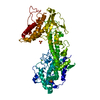
| Entry | Database: PDB / ID: 6rgo | ||||||
|---|---|---|---|---|---|---|---|
| Title | Complex of KlAtg21 with coiled-coil of AgAtg16 | ||||||
 Components Components |
| ||||||
 Keywords Keywords | LIPID BINDING PROTEIN /  Autophagy / yeast / Atg16 / Atg21 / Atg8 lipidation Autophagy / yeast / Atg16 / Atg21 / Atg8 lipidation | ||||||
| Function / homology |  Function and homology information Function and homology information: / : / Atg8-family ligase activity / Atg12-Atg5-Atg16 complex / C-terminal protein lipidation / phagophore / protein lipidation / vacuole-isolation membrane contact site / cytoplasm to vacuole targeting by the Cvt pathway / vesicle organization ...: / : / Atg8-family ligase activity / Atg12-Atg5-Atg16 complex / C-terminal protein lipidation / phagophore / protein lipidation / vacuole-isolation membrane contact site / cytoplasm to vacuole targeting by the Cvt pathway / vesicle organization / autophagosome organization / autophagy of mitochondrion / protein localization to phagophore assembly site / phagophore assembly site membrane / piecemeal microautophagy of the nucleus / phosphatidylinositol-3-phosphate binding / fungal-type vacuole membrane / phagophore assembly site / phosphatidylinositol-4-phosphate binding / phosphatidylinositol-3,5-bisphosphate binding / protein-macromolecule adaptor activity / endosome / identical protein binding / cytosolSimilarity search - Function | ||||||
| Biological species |  Kluyveromyces lactis (yeast) Kluyveromyces lactis (yeast) Ashbya gossypii (fungus) Ashbya gossypii (fungus) | ||||||
| Method | X-RAY DIFFRACTION / SYNCHROTRON / MOLECULAR REPLACEMENT / molecular replacement / Resolution: 3.701 Å | ||||||
 Authors Authors | Thumm, M. / Neumann, P. | ||||||
| Funding support |  Germany, 1items Germany, 1items
| ||||||
 Citation Citation | Journal: Autophagy / Year: 2020 Title: Atg21 organizes Atg8 lipidation at the contact of the vacuole with the phagophore. Authors: Munzel, L. / Neumann, P. / Otto, F.B. / Krick, R. / Metje-Sprink, J. / Kroppen, B. / Karedla, N. / Enderlein, J. / Meinecke, M. / Ficner, R. / Thumm, M. | ||||||
| History |
|
- Structure visualization
| Structure viewer | Molecule: MolmilJmol/JSmol |
|---|
- Downloads & links
-Download
| PDBx/mmCIF format | 6rgo.cif.gz | 297.2 KB | Display | PDBx/mmCIF format |
|---|---|---|---|---|
| PDB format | pdb6rgo.ent.gz | 246.1 KB | Display | PDB format |
| PDBx/mmJSON format | 6rgo.json.gz | Tree view | PDBx/mmJSON format | |
| Others |  Other downloads Other downloads |
-Validation report
| Arichive directory | https://data.pdbj.org/pub/pdb/validation_reports/rg/6rgoftp://data.pdbj.org/pub/pdb/validation_reports/rg/6rgo | HTTPS FTP |
|---|
-Related structure data
| Related structure data |  5ltgS S: Starting model for refinement |
|---|---|
| Similar structure data |


-Links
 PDBj
PDBj
- Assembly
| Deposited unit | 
| ||||||||
|---|---|---|---|---|---|---|---|---|---|
| 1 |
| ||||||||
| Unit cell |
|
-Components
| #1: Protein | Mass: 43607.934 Da / Num. of mol.: 2 Source method: isolated from a genetically manipulated source Source: (gene. exp.) Kluyveromyces lactis (strain ATCC 8585 / CBS 2359 / DSM 70799 / NBRC 1267 / NRRL Y-1140 / WM37) (yeast)Strain: ATCC 8585 / CBS 2359 / DSM 70799 / NBRC 1267 / NRRL Y-1140 / WM37 Gene: ATG21, KLLA0E24354g / Production host:  Escherichia coli (E. coli) / References: UniProt: Q6CLZ2 Escherichia coli (E. coli) / References: UniProt: Q6CLZ2#2: Protein | Mass: 6165.890 Da / Num. of mol.: 2 Source method: isolated from a genetically manipulated source Source: (gene. exp.) Ashbya gossypii (strain ATCC 10895 / CBS 109.51 / FGSC 9923 / NRRL Y-1056) (fungus)Strain: ATCC 10895 / CBS 109.51 / FGSC 9923 / NRRL Y-1056 / Gene: ATG16, AFL186W / Production host: Escherichia coli (E. coli) / References: UniProt: Q755K3 |
|---|
-Experimental details
-Experiment
| Experiment | Method: X-RAY DIFFRACTION / Number of used crystals: 1 |
|---|
- Sample preparation
| Crystal | Density Matthews: 4.09 Å3/Da / Density % sol: 69.94 % |
|---|---|
| Crystal grow | Temperature: 293.15 K / Method: vapor diffusion, hanging drop / pH: 7.5 Details: Protein concetration 13 mg/mL, Clostripain for in situ proteolysis (1:2000 ratio). Reservoir: ProComplex (Molecular Dimension) B11: 15 % PEG4000, 0.1 M HEPES pH 7.5. Droplet: 2 microL ...Details: Protein concetration 13 mg/mL, Clostripain for in situ proteolysis (1:2000 ratio). Reservoir: ProComplex (Molecular Dimension) B11: 15 % PEG4000, 0.1 M HEPES pH 7.5. Droplet: 2 microL Protein (mit 1:2000 Clostripain) + 3 microL Reservoir. |
-Data collection
| Diffraction | Mean temperature: 100 K / Serial crystal experiment: N | ||||||||||||||||||||||||||||||
|---|---|---|---|---|---|---|---|---|---|---|---|---|---|---|---|---|---|---|---|---|---|---|---|---|---|---|---|---|---|---|---|
| Diffraction source | Source: SYNCHROTRON / Site: SLS  / Beamline: X06SA / Wavelength: 0.97918 Å / Beamline: X06SA / Wavelength: 0.97918 Å | ||||||||||||||||||||||||||||||
| Detector | Type: DECTRIS PILATUS 6M / Detector: PIXEL / Date: Aug 8, 2016 | ||||||||||||||||||||||||||||||
| Radiation | Protocol: SINGLE WAVELENGTH / Monochromatic (M) / Laue (L): M / Scattering type: x-ray | ||||||||||||||||||||||||||||||
| Radiation wavelength | Wavelength: 0.97918 Å / Relative weight: 1 | ||||||||||||||||||||||||||||||
| Reflection | Resolution: 3.7→46.29 Å / Num. obs: 17917 / % possible obs: 99.6 % / Redundancy: 14.2 % / CC1/2: 0.999 / Rmerge(I) obs: 0.23 / Rpim(I) all: 0.062 / Rrim(I) all: 0.239 / Net I/σ(I): 9.4 / Num. measured all: 254489 / Scaling rejects: 9 | ||||||||||||||||||||||||||||||
| Reflection shell | Diffraction-ID: 1
|
-Phasing
| Phasing | Method: molecular replacement |
|---|
- Processing
| Software |
| |||||||||||||||||||||||||||||||||||||||||||||||||||||||||||||||||||||||||||||||||||||||||||||||||||||||||||||||||||||||||||||
|---|---|---|---|---|---|---|---|---|---|---|---|---|---|---|---|---|---|---|---|---|---|---|---|---|---|---|---|---|---|---|---|---|---|---|---|---|---|---|---|---|---|---|---|---|---|---|---|---|---|---|---|---|---|---|---|---|---|---|---|---|---|---|---|---|---|---|---|---|---|---|---|---|---|---|---|---|---|---|---|---|---|---|---|---|---|---|---|---|---|---|---|---|---|---|---|---|---|---|---|---|---|---|---|---|---|---|---|---|---|---|---|---|---|---|---|---|---|---|---|---|---|---|---|---|---|---|
| Refinement | Method to determine structure: MOLECULAR REPLACEMENT Starting model: 5LTG Resolution: 3.701→46.29 Å / SU ML: 0.65 / Cross valid method: THROUGHOUT / σ(F): 0.41 / Phase error: 34.59
| |||||||||||||||||||||||||||||||||||||||||||||||||||||||||||||||||||||||||||||||||||||||||||||||||||||||||||||||||||||||||||||
| Solvent computation | Shrinkage radii: 0.8 Å / VDW probe radii: 1.1 Å | |||||||||||||||||||||||||||||||||||||||||||||||||||||||||||||||||||||||||||||||||||||||||||||||||||||||||||||||||||||||||||||
| Refinement step | Cycle: LAST / Resolution: 3.701→46.29 Å
| |||||||||||||||||||||||||||||||||||||||||||||||||||||||||||||||||||||||||||||||||||||||||||||||||||||||||||||||||||||||||||||
| Refine LS restraints |
| |||||||||||||||||||||||||||||||||||||||||||||||||||||||||||||||||||||||||||||||||||||||||||||||||||||||||||||||||||||||||||||
| LS refinement shell |
| |||||||||||||||||||||||||||||||||||||||||||||||||||||||||||||||||||||||||||||||||||||||||||||||||||||||||||||||||||||||||||||
| Refinement TLS params. | Method: refined / Refine-ID: X-RAY DIFFRACTION
| |||||||||||||||||||||||||||||||||||||||||||||||||||||||||||||||||||||||||||||||||||||||||||||||||||||||||||||||||||||||||||||
| Refinement TLS group |
|
