 Movie
Movie Controller

[English] 日本語
 Yorodumi
- PDB-3hw9: Cation selective pathway of OmpF porin revealed by anomalous x-ra... -
+ Open data

- Basic information
| Entry | Database: PDB / ID: 3hw9 | ||||||
|---|---|---|---|---|---|---|---|
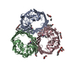
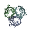
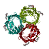
| Title | Cation selective pathway of OmpF porin revealed by anomalous x-ray diffraction | ||||||
 Components Components | Outer membrane protein F | ||||||
 Keywords Keywords |  MEMBRANE PROTEIN / ion transport / porin / integral membrane protein porin / Cell membrane / Cell outer membrane / Membrane / Phage recognition / Transmembrane / Transport MEMBRANE PROTEIN / ion transport / porin / integral membrane protein porin / Cell membrane / Cell outer membrane / Membrane / Phage recognition / Transmembrane / Transport | ||||||
| Function / homology |  Function and homology information Function and homology informationcolicin transmembrane transporter activity / monoatomic ion channel complex / porin activity / pore complex / protein homotrimerization / monoatomic ion transmembrane transport / lipopolysaccharide binding / cell outer membrane / disordered domain specific binding / protein transport ...colicin transmembrane transporter activity / monoatomic ion channel complex / porin activity / pore complex / protein homotrimerization / monoatomic ion transmembrane transport / lipopolysaccharide binding / cell outer membrane / disordered domain specific binding / protein transport / monoatomic ion channel activity / lipid binding / membrane / identical protein bindingSimilarity search - Function | ||||||
| Biological species |  Escherichia coli (E. coli) Escherichia coli (E. coli) | ||||||
| Method | X-RAY DIFFRACTION / SYNCHROTRON / MOLECULAR REPLACEMENT / Resolution: 2.61 Å | ||||||
 Authors Authors | Balasundaresan, D. / Raychaudhury, S. / Blachowicz, L. / Roux, B. | ||||||
 Citation Citation | Journal: J.Mol.Biol. / Year: 2010 Title: Cation-selective pathway of OmpF porin revealed by anomalous X-ray diffraction. Authors: Dhakshnamoorthy, B. / Raychaudhury, S. / Blachowicz, L. / Roux, B. | ||||||
| History |
|
- Structure visualization
| Structure viewer | Molecule: MolmilJmol/JSmol |
|---|
- Downloads & links
-Download
| PDBx/mmCIF format | 3hw9.cif.gz | 145.3 KB | Display | PDBx/mmCIF format |
|---|---|---|---|---|
| PDB format | pdb3hw9.ent.gz | 113.1 KB | Display | PDB format |
| PDBx/mmJSON format | 3hw9.json.gz | Tree view | PDBx/mmJSON format | |
| Others |  Other downloads Other downloads |
-Validation report
| Arichive directory | https://data.pdbj.org/pub/pdb/validation_reports/hw/3hw9ftp://data.pdbj.org/pub/pdb/validation_reports/hw/3hw9 | HTTPS FTP |
|---|
-Related structure data
| Related structure data |  3hwbC  2omfS C: citing same article ( S: Starting model for refinement |
|---|---|
| Similar structure data |


-Links
 PDBj
PDBj- Assembly
| Deposited unit | 
| ||||||||
|---|---|---|---|---|---|---|---|---|---|
| 1 | 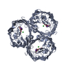
| ||||||||
| 2 | 
| ||||||||
| 3 |
| ||||||||
| Unit cell |
|
-Components
| #1: Protein | Mass: 39365.043 Da / Num. of mol.: 2 Source method: isolated from a genetically manipulated source Source: (gene. exp.) Escherichia coli (E. coli) / Strain: K-12 / Gene: b0929, cmlB, coa, cry, JW0912, ompF, tolF / Plasmid: pPR272 / Production host: Escherichia coli (E. coli) / Strain (production host): MH225 / References: UniProt: P02931#2: Chemical | Polyethylene glycol  Mass: 282.331 Da / Num. of mol.: 2 / Source method: obtained synthetically / Formula: C12H26O7 / Comment: precipitant*YM Mass: 282.331 Da / Num. of mol.: 2 / Source method: obtained synthetically / Formula: C12H26O7 / Comment: precipitant*YM#3: Chemical | ChemComp-K /   Mass: 39.098 Da / Num. of mol.: 4 / Source method: obtained synthetically / Formula: K Mass: 39.098 Da / Num. of mol.: 4 / Source method: obtained synthetically / Formula: K#4: Chemical | Chloride  Mass: 35.453 Da / Num. of mol.: 2 / Source method: obtained synthetically / Formula: Cl Mass: 35.453 Da / Num. of mol.: 2 / Source method: obtained synthetically / Formula: Cl#5: Water | ChemComp-HOH / | Water Mass: 18.015 Da / Num. of mol.: 134 / Source method: isolated from a natural source / Formula: H2O Mass: 18.015 Da / Num. of mol.: 134 / Source method: isolated from a natural source / Formula: H2O |
|---|
-Experimental details
-Experiment
| Experiment | Method: X-RAY DIFFRACTION / Number of used crystals: 1 |
|---|
- Sample preparation
| Crystal | Density Matthews: 3.07 Å3/Da / Density % sol: 59.96 % |
|---|---|
| Crystal grow | Temperature: 293 K / Method: vapor diffusion, sitting drop / pH: 6.5 Details: 0.5M NaCl, Na Phosphate pH 6.5, 0.9% BOG, 0.09% octylPOE and 14-18% PEG2000, VAPOR DIFFUSION, SITTING DROP, temperature 293K |
-Data collection
| Diffraction | Mean temperature: 100 K |
|---|---|
| Diffraction source | Source: SYNCHROTRON / Site: APS  / Beamline: 23-ID-B / Wavelength: 1.033 Å / Beamline: 23-ID-B / Wavelength: 1.033 Å |
| Detector | Type: ADSC QUANTUM 315 / Detector: CCD / Date: Jun 8, 2007 |
| Radiation | Monochromator: Si(111) channel / Protocol: SINGLE WAVELENGTH / Monochromatic (M) / Laue (L): M / Scattering type: x-ray |
| Radiation wavelength | Wavelength: 1.033 Å / Relative weight: 1 |
| Reflection | Resolution: 2.61→50 Å / Num. obs: 26020 / % possible obs: 89.2 % / Observed criterion σ(F): 0 / Observed criterion σ(I): 0 / Redundancy: 6.8 % / Rmerge(I) obs: 0.147 / Net I/σ(I): 28.9 |
| Reflection shell | Resolution: 2.61→2.69 Å / Redundancy: 4 % / Rmerge(I) obs: 0.4 / Mean I/σ(I) obs: 5.51 / Num. unique all: 2154 / % possible all: 74.5 |
- Processing
| Software |
| ||||||||||||||||||||||||||||||||||||||||||||||||||||||||||||||||||||||||||||||||||||||||||||||||||||||||||||||||||||||||||||||||||||||||||||||||||||||||||||||||||||||||||
|---|---|---|---|---|---|---|---|---|---|---|---|---|---|---|---|---|---|---|---|---|---|---|---|---|---|---|---|---|---|---|---|---|---|---|---|---|---|---|---|---|---|---|---|---|---|---|---|---|---|---|---|---|---|---|---|---|---|---|---|---|---|---|---|---|---|---|---|---|---|---|---|---|---|---|---|---|---|---|---|---|---|---|---|---|---|---|---|---|---|---|---|---|---|---|---|---|---|---|---|---|---|---|---|---|---|---|---|---|---|---|---|---|---|---|---|---|---|---|---|---|---|---|---|---|---|---|---|---|---|---|---|---|---|---|---|---|---|---|---|---|---|---|---|---|---|---|---|---|---|---|---|---|---|---|---|---|---|---|---|---|---|---|---|---|---|---|---|---|---|---|---|
| Refinement | Method to determine structure: MOLECULAR REPLACEMENT Starting model: PDB entry 2OMF Resolution: 2.61→46.93 Å / Cor.coef. Fo:Fc: 0.907 / Cor.coef. Fo:Fc free: 0.847 / SU B: 12.978 / SU ML: 0.285 / Cross valid method: THROUGHOUT / ESU R: 0.877 / ESU R Free: 0.39 / Stereochemistry target values: MAXIMUM LIKELIHOOD
| ||||||||||||||||||||||||||||||||||||||||||||||||||||||||||||||||||||||||||||||||||||||||||||||||||||||||||||||||||||||||||||||||||||||||||||||||||||||||||||||||||||||||||
| Solvent computation | Ion probe radii: 0.8 Å / Shrinkage radii: 0.8 Å / VDW probe radii: 1.2 Å / Solvent model: MASK | ||||||||||||||||||||||||||||||||||||||||||||||||||||||||||||||||||||||||||||||||||||||||||||||||||||||||||||||||||||||||||||||||||||||||||||||||||||||||||||||||||||||||||
| Displacement parameters | Biso mean: 51.921 Å2
| ||||||||||||||||||||||||||||||||||||||||||||||||||||||||||||||||||||||||||||||||||||||||||||||||||||||||||||||||||||||||||||||||||||||||||||||||||||||||||||||||||||||||||
| Refinement step | Cycle: LAST / Resolution: 2.61→46.93 Å
| ||||||||||||||||||||||||||||||||||||||||||||||||||||||||||||||||||||||||||||||||||||||||||||||||||||||||||||||||||||||||||||||||||||||||||||||||||||||||||||||||||||||||||
| Refine LS restraints |
| ||||||||||||||||||||||||||||||||||||||||||||||||||||||||||||||||||||||||||||||||||||||||||||||||||||||||||||||||||||||||||||||||||||||||||||||||||||||||||||||||||||||||||
| LS refinement shell | Resolution: 2.61→2.678 Å / Total num. of bins used: 20
|
