 Movie
Movie Controller

[English] 日本語
 Yorodumi
- PDB-6pzq: Structure of the human respiratory syncytial virus M2-1 protein i... -
+ Open data

- Basic information
| Entry | Database: PDB / ID: 6pzq | ||||||
|---|---|---|---|---|---|---|---|
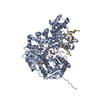
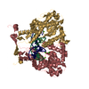
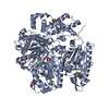
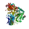
| Title | Structure of the human respiratory syncytial virus M2-1 protein in complex with a short positive-sense gene-end RNA | ||||||
 Components Components |
| ||||||
 Keywords Keywords | METAL BINDING PROTEIN/RNA / HRSV M2-1 RNA complex /  RNA BINDING PROTEIN / METAL BINDING PROTEIN-RNA complex RNA BINDING PROTEIN / METAL BINDING PROTEIN-RNA complex | ||||||
| Function / homology |  Function and homology information Function and homology information: / regulation of viral transcription / viral transcription / transcription antitermination / virion component / host cell cytoplasm / host cell nucleus / structural molecule activity / RNA binding / metal ion bindingSimilarity search - Function | ||||||
| Biological species |  Human respiratory syncytial virus AHuman respiratory syncytial virus A2 Human respiratory syncytial virus AHuman respiratory syncytial virus A2 | ||||||
| Method | X-RAY DIFFRACTION / SYNCHROTRON / MOLECULAR REPLACEMENT / molecular replacement / Resolution: 2.703 Å | ||||||
 Authors Authors | Gao, Y. / Cao, D. / Liang, B. | ||||||
| Funding support |  United States, 1items United States, 1items
| ||||||
 Citation Citation | Journal: Structure / Year: 2020 Title: Structure of the Human Respiratory Syncytial Virus M2-1 Protein in Complex with a Short Positive-Sense Gene-End RNA. Authors: Gao, Y. / Cao, D. / Pawnikar, S. / John, K.P. / Ahn, H.M. / Hill, S. / Ha, J.M. / Parikh, P. / Ogilvie, C. / Swain, A. / Yang, A. / Bell, A. / Salazar, A. / Miao, Y. / Liang, B. | ||||||
| History |
|
- Structure visualization
| Structure viewer | Molecule: MolmilJmol/JSmol |
|---|
- Downloads & links
-Download
| PDBx/mmCIF format | 6pzq.cif.gz | 263.4 KB | Display | PDBx/mmCIF format |
|---|---|---|---|---|
| PDB format | pdb6pzq.ent.gz | 212.9 KB | Display | PDB format |
| PDBx/mmJSON format | 6pzq.json.gz | Tree view | PDBx/mmJSON format | |
| Others |  Other downloads Other downloads |
-Validation report
| Arichive directory | https://data.pdbj.org/pub/pdb/validation_reports/pz/6pzqftp://data.pdbj.org/pub/pdb/validation_reports/pz/6pzq | HTTPS FTP |
|---|
-Related structure data
| Related structure data |  4c3bS S: Starting model for refinement |
|---|---|
| Similar structure data |


-Links
 PDBj
PDBj
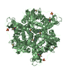
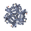
- Assembly
| Deposited unit | 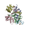
| ||||||||
|---|---|---|---|---|---|---|---|---|---|
| 1 |
| ||||||||
| Unit cell |
|
-Components
| #1: Protein | Mass: 22216.404 Da / Num. of mol.: 4 Source method: isolated from a genetically manipulated source Source: (gene. exp.) Human respiratory syncytial virus A (strain A2)Strain: A2 / Gene: M2-1 / Production host:  Escherichia coli (E. coli) / References: UniProt: P04545 Escherichia coli (E. coli) / References: UniProt: P04545#2: RNA chain | Mass: 2206.363 Da / Num. of mol.: 2 / Source method: obtained synthetically / Source: (synth.) Human respiratory syncytial virus A2#3: Chemical | ChemComp-ZN /   Mass: 65.409 Da / Num. of mol.: 4 / Source method: obtained synthetically / Formula: Zn Mass: 65.409 Da / Num. of mol.: 4 / Source method: obtained synthetically / Formula: Zn#4: Water | ChemComp-HOH / | Water Mass: 18.015 Da / Num. of mol.: 14 / Source method: isolated from a natural source / Formula: H2O Mass: 18.015 Da / Num. of mol.: 14 / Source method: isolated from a natural source / Formula: H2OHas ligand of interest | N | |
|---|
-Experimental details
-Experiment
| Experiment | Method: X-RAY DIFFRACTION / Number of used crystals: 1 |
|---|
- Sample preparation
| Crystal | Density Matthews: 2.57 Å3/Da / Density % sol: 52.1 % |
|---|---|
| Crystal grow | Temperature: 289 K / Method: vapor diffusion, hanging drop Details: 0.2 M magnesium chloride hexahydrate, 0.1 M Tris pH 8.5, 25% w/v polyethylene glycol 3,350 |
-Data collection
| Diffraction | Mean temperature: 100 K / Serial crystal experiment: N |
|---|---|
| Diffraction source | Source: SYNCHROTRON / Site: APS / Beamline: 22-ID / Wavelength: 1 Å |
| Detector | Type: MAR CCD 130 mm / Detector: CCD / Date: Apr 15, 2018 |
| Radiation | Protocol: SINGLE WAVELENGTH / Monochromatic (M) / Laue (L): M / Scattering type: x-ray |
| Radiation wavelength | Wavelength: 1 Å / Relative weight: 1 |
| Reflection | Resolution: 2.703→94.75 Å / Num. obs: 25701 / % possible obs: 99.8 % / Redundancy: 24.3 % / Biso Wilson estimate: 89.84 Å2 / CC1/2: 0.998 / Rmerge(I) obs: 0.127 / Rpim(I) all: 0.027 / Rrim(I) all: 0.132 / Net I/σ(I): 13.3 |
| Reflection shell | Resolution: 2.703→2.85 Å / Redundancy: 26 % / Num. unique obs: 3673 / Rpim(I) all: 0.39 / % possible all: 100 |
-Phasing
| Phasing | Method: molecular replacement |
|---|
- Processing
| Software |
| ||||||||||||||||||||||||||||||||||||||||||||||||||||||||||||
|---|---|---|---|---|---|---|---|---|---|---|---|---|---|---|---|---|---|---|---|---|---|---|---|---|---|---|---|---|---|---|---|---|---|---|---|---|---|---|---|---|---|---|---|---|---|---|---|---|---|---|---|---|---|---|---|---|---|---|---|---|---|
| Refinement | Method to determine structure: MOLECULAR REPLACEMENT Starting model: 4C3B Resolution: 2.703→63.517 Å / SU ML: 0.44 / Cross valid method: THROUGHOUT / σ(F): 1.33 / Phase error: 33.07 / Stereochemistry target values: ML
| ||||||||||||||||||||||||||||||||||||||||||||||||||||||||||||
| Solvent computation | Shrinkage radii: 0.9 Å / VDW probe radii: 1.11 Å / Solvent model: FLAT BULK SOLVENT MODEL | ||||||||||||||||||||||||||||||||||||||||||||||||||||||||||||
| Displacement parameters | Biso max: 211.15 Å2 / Biso mean: 104.4578 Å2 / Biso min: 36.64 Å2 | ||||||||||||||||||||||||||||||||||||||||||||||||||||||||||||
| Refinement step | Cycle: final / Resolution: 2.703→63.517 Å
| ||||||||||||||||||||||||||||||||||||||||||||||||||||||||||||
| Refine LS restraints |
| ||||||||||||||||||||||||||||||||||||||||||||||||||||||||||||
| LS refinement shell | Refine-ID: X-RAY DIFFRACTION / Rfactor Rfree error: 0
|
