 Movie
Movie Controller

[English] 日本語
 Yorodumi
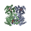
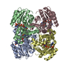
- PDB-4bge: Crystal structure of InhA(S94A) mutant in complex with pyridomycin -
+ Open data

- Basic information
| Entry | Database: PDB / ID: 4bge | ||||||
|---|---|---|---|---|---|---|---|
| Title | Crystal structure of InhA(S94A) mutant in complex with pyridomycin | ||||||
 Components Components | Enoyl-[acyl-carrier-protein] reductase [NADH] | ||||||
 Keywords Keywords |  OXIDOREDUCTASE OXIDOREDUCTASE | ||||||
| Function / homology |  Function and homology information Function and homology informationtrans-2-enoyl-CoA reductase (NADH) activity / mycolic acid biosynthetic process / fatty acid elongation / enoyl-[acyl-carrier-protein] reductase (NADH) / enoyl-[acyl-carrier-protein] reductase (NADH) activity / NAD+ binding / peptidoglycan-based cell wall / fatty acid binding / response to antibiotic / plasma membraneSimilarity search - Function | ||||||
| Biological species |   Mycobacterium tuberculosis (bacteria) Mycobacterium tuberculosis (bacteria) | ||||||
| Method | X-RAY DIFFRACTION / SYNCHROTRON / MOLECULAR REPLACEMENT / Resolution: 2.25 Å | ||||||
 Authors Authors | Pojer, F. / Hartkoorn, R.C. / Cole, S.T. | ||||||
 Citation Citation | Journal: Nat. Chem. Biol. / Year: 2014 Title: Pyridomycin bridges the NADH- and substrate-binding pockets of the enoyl reductase InhA. Authors: Hartkoorn, R.C. / Pojer, F. / Read, J.A. / Gingell, H. / Neres, J. / Horlacher, O.P. / Altmann, K.H. / Cole, S.T. | ||||||
| History |
|
- Structure visualization
| Structure viewer | Molecule: MolmilJmol/JSmol |
|---|
- Downloads & links
-Download
| PDBx/mmCIF format | 4bge.cif.gz | 314.5 KB | Display | PDBx/mmCIF format |
|---|---|---|---|---|
| PDB format | pdb4bge.ent.gz | 257.2 KB | Display | PDB format |
| PDBx/mmJSON format | 4bge.json.gz | Tree view | PDBx/mmJSON format | |
| Others |  Other downloads Other downloads |
-Validation report
| Arichive directory | https://data.pdbj.org/pub/pdb/validation_reports/bg/4bgeftp://data.pdbj.org/pub/pdb/validation_reports/bg/4bge | HTTPS FTP |
|---|
-Related structure data
| Related structure data |  4bgiC 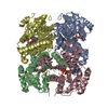 4biiC  4dtiS C: citing same article ( S: Starting model for refinement |
|---|---|
| Similar structure data |


-Links
 PDBj
PDBj

- Assembly
| Deposited unit | 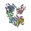
| ||||||||
|---|---|---|---|---|---|---|---|---|---|
| 1 | 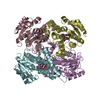
| ||||||||
| 2 | 
| ||||||||
| 3 | 
| ||||||||
| Unit cell |
|
-Components
| #1: Protein | Mass: 28538.781 Da / Num. of mol.: 6 / Mutation: S94A Source method: isolated from a genetically manipulated source Source: (gene. exp.) Mycobacterium tuberculosis (strain ATCC 25618 / H37Rv) (bacteria)Gene: inhA, Rv1484, MTCY277.05 / Production host: Escherichia coli BL21(DE3) (bacteria)References: UniProt: P9WGR1, enoyl-[acyl-carrier-protein] reductase (NADH) #2: Chemical | ChemComp-PYW /   Mass: 540.565 Da / Num. of mol.: 6 / Source method: obtained synthetically / Formula: C27H32N4O8 Mass: 540.565 Da / Num. of mol.: 6 / Source method: obtained synthetically / Formula: C27H32N4O8#3: Water | ChemComp-HOH / | Water Mass: 18.015 Da / Num. of mol.: 727 / Source method: isolated from a natural source / Formula: H2O Mass: 18.015 Da / Num. of mol.: 727 / Source method: isolated from a natural source / Formula: H2O |
|---|
-Experimental details
-Experiment
| Experiment | Method: X-RAY DIFFRACTION / Number of used crystals: 1 |
|---|
- Sample preparation
| Crystal | Density Matthews: 2.32 Å3/Da / Density % sol: 46.93 % / Description: NONE |
|---|
-Data collection
| Diffraction | Mean temperature: 100 K |
|---|---|
| Diffraction source | Source: SYNCHROTRON / Site: SLS  / Beamline: X06SA / Wavelength: 1 / Beamline: X06SA / Wavelength: 1 |
| Detector | Date: Dec 16, 2012 |
| Radiation | Protocol: SINGLE WAVELENGTH / Monochromatic (M) / Laue (L): M / Scattering type: x-ray |
| Radiation wavelength | Wavelength: 1 Å / Relative weight: 1 |
| Reflection | Resolution: 2.25→50 Å / Num. obs: 140935 / % possible obs: 96.2 % / Observed criterion σ(I): 0 / Redundancy: 3 % / Biso Wilson estimate: 31.05 Å2 / Rrim(I) all: 0.141 / Net I/σ(I): 8.5 |
| Reflection shell | Resolution: 2.25→2.38 Å / Redundancy: 3 % / Mean I/σ(I) obs: 2.6 / Rrim(I) all: 0.592 / % possible all: 91.7 |
- Processing
| Software |
| ||||||||||||||||||||||||||||||||||||||||||||||||||||||||||||||||||||||||||||||||||||||||||||||||||||||||||||||||||
|---|---|---|---|---|---|---|---|---|---|---|---|---|---|---|---|---|---|---|---|---|---|---|---|---|---|---|---|---|---|---|---|---|---|---|---|---|---|---|---|---|---|---|---|---|---|---|---|---|---|---|---|---|---|---|---|---|---|---|---|---|---|---|---|---|---|---|---|---|---|---|---|---|---|---|---|---|---|---|---|---|---|---|---|---|---|---|---|---|---|---|---|---|---|---|---|---|---|---|---|---|---|---|---|---|---|---|---|---|---|---|---|---|---|---|---|
| Refinement | Method to determine structure: MOLECULAR REPLACEMENT Starting model: PDB ENTRY 4DTI Resolution: 2.25→47.5 Å / Cor.coef. Fo:Fc: 0.8173 / Cor.coef. Fo:Fc free: 0.7606 / SU R Cruickshank DPI: 0.395 / Cross valid method: THROUGHOUT / σ(F): 0 / SU R Blow DPI: 0.399 / SU Rfree Blow DPI: 0.256 / SU Rfree Cruickshank DPI: 0.258
| ||||||||||||||||||||||||||||||||||||||||||||||||||||||||||||||||||||||||||||||||||||||||||||||||||||||||||||||||||
| Displacement parameters | Biso mean: 33.58 Å2
| ||||||||||||||||||||||||||||||||||||||||||||||||||||||||||||||||||||||||||||||||||||||||||||||||||||||||||||||||||
| Refine analyze | Luzzati coordinate error obs: 0.487 Å | ||||||||||||||||||||||||||||||||||||||||||||||||||||||||||||||||||||||||||||||||||||||||||||||||||||||||||||||||||
| Refinement step | Cycle: LAST / Resolution: 2.25→47.5 Å
| ||||||||||||||||||||||||||||||||||||||||||||||||||||||||||||||||||||||||||||||||||||||||||||||||||||||||||||||||||
| Refine LS restraints |
| ||||||||||||||||||||||||||||||||||||||||||||||||||||||||||||||||||||||||||||||||||||||||||||||||||||||||||||||||||
| LS refinement shell | Resolution: 2.25→2.31 Å / Total num. of bins used: 20
|
