 Movie
Movie Controller

[English] 日本語
 Yorodumi
- PDB-6psk: Crystal structure of the complex between periplasmic domains of a... -
+ Open data

- Basic information
| Entry | Database: PDB / ID: 6psk | ||||||
|---|---|---|---|---|---|---|---|
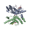
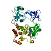
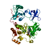
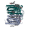
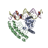
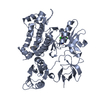
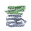
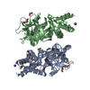
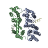
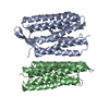
| Title | Crystal structure of the complex between periplasmic domains of antiholin RI and holin T from T4 phage, in P6522 | ||||||
 Components Components |
| ||||||
 Keywords Keywords |  VIRAL PROTEIN / phage / lysis inhibition VIRAL PROTEIN / phage / lysis inhibition | ||||||
| Function / homology |  Function and homology information Function and homology information: / host cell periplasmic space / pore-forming activity / cytolysis / molecular function inhibitor activity / viral release from host cell by cytolysis / killing of cells of another organism / host cell plasma membrane / DNA binding / membraneSimilarity search - Function | ||||||
| Biological species |  Escherichia phage ECML-134 (virus)Escherichia phage vB_EcoM_NBG2 (virus) Escherichia phage ECML-134 (virus)Escherichia phage vB_EcoM_NBG2 (virus) | ||||||
| Method | X-RAY DIFFRACTION / SYNCHROTRON / SAD / Resolution: 2.2 Å | ||||||
 Authors Authors | Kuznetsov, V.B. / Krieger, I.V. / Sacchettini, J.C. | ||||||
| Funding support |  United States, 1items United States, 1items
| ||||||
 Citation Citation | Journal: J Mol Biol / Year: 2020 Title: The Structural Basis of T4 Phage Lysis Control: DNA as the Signal for Lysis Inhibition. Authors: Inna V Krieger / Vladimir Kuznetsov / Jeng-Yih Chang / Junjie Zhang / Samir H Moussa / Ryland F Young / James C Sacchettini / Abstract: Optimal phage propagation depends on the regulation of the lysis of the infected host cell. In T4 phage infection, lysis occurs when the holin protein (T) forms lesions in the host membrane. However, ...Optimal phage propagation depends on the regulation of the lysis of the infected host cell. In T4 phage infection, lysis occurs when the holin protein (T) forms lesions in the host membrane. However, the lethal function of T can be blocked by an antiholin (RI) during lysis inhibition (LIN). LIN sets if the infected cell undergoes superinfection, then the lysis is delayed until host/phage ratio becomes more favorable for the release of progeny. It has been thought that a signal derived from the superinfection is required to activate RI. Here we report structures that suggest a radically different model in which RI binds to T irrespective of superinfection, causing it to accumulate in a membrane as heterotetrameric 2RI-2T complex. Moreover, we show the complex binds non-specifically to DNA, suggesting that the gDNA from the superinfecting phage serves as the LIN signal and that stabilization of the complex by DNA binding is what defines LIN. Finally, we show that soluble domain of free RI crystallizes in a domain-swapped homotetramer, which likely works as a sink for RI molecules released from the RI-T complex to ensure efficient lysis. These results constitute the first structural basis and a new model not only for the historic LIN phenomenon but also for the temporal regulation of phage lysis in general. | ||||||
| History |
|
- Structure visualization
| Structure viewer | Molecule: MolmilJmol/JSmol |
|---|
- Downloads & links
-Download
| PDBx/mmCIF format | 6psk.cif.gz | 59.2 KB | Display | PDBx/mmCIF format |
|---|---|---|---|---|
| PDB format | pdb6psk.ent.gz | 45.2 KB | Display | PDB format |
| PDBx/mmJSON format | 6psk.json.gz | Tree view | PDBx/mmJSON format | |
| Others |  Other downloads Other downloads |
-Validation report
| Arichive directory | https://data.pdbj.org/pub/pdb/validation_reports/ps/6pskftp://data.pdbj.org/pub/pdb/validation_reports/ps/6psk | HTTPS FTP |
|---|
-Related structure data
-Links
 PDBj
PDBj- Assembly
| Deposited unit | 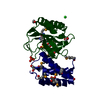
| |||||||||
|---|---|---|---|---|---|---|---|---|---|---|
| 1 | 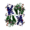
| |||||||||
| Unit cell |
| |||||||||
| Components on special symmetry positions |
|
-Components
-Protein , 2 types, 2 molecules RT
| #1: Protein | Mass: 9083.254 Da / Num. of mol.: 1 Source method: isolated from a genetically manipulated source Source: (gene. exp.) Escherichia phage ECML-134 (virus) / Gene: ECML134_104 / Production host:  Escherichia coli (E. coli) / References: UniProt: I7AU04, UniProt: P13304*PLUS Escherichia coli (E. coli) / References: UniProt: I7AU04, UniProt: P13304*PLUS |
|---|---|
| #2: Protein | Mass: 18102.947 Da / Num. of mol.: 1 Source method: isolated from a genetically manipulated source Source: (gene. exp.) Escherichia phage vB_EcoM_NBG2 (virus) / Gene: vBEcoMNBG2_239 / Production host: Escherichia coli (E. coli) / References: UniProt: A0A2U8QQK7, UniProt: P06808*PLUS |
-Non-polymers , 5 types, 82 molecules 


| #3: Chemical | Sulfate Mass: 96.063 Da / Num. of mol.: 3 / Source method: isolated from a natural source / Formula: SO4 Mass: 96.063 Da / Num. of mol.: 3 / Source method: isolated from a natural source / Formula: SO4#4: Chemical | ChemComp-EDO / Ethylene glycol Mass: 62.068 Da / Num. of mol.: 4 / Source method: obtained synthetically / Formula: C2H6O2 Mass: 62.068 Da / Num. of mol.: 4 / Source method: obtained synthetically / Formula: C2H6O2#5: Chemical | ChemComp-BTB / | Bis-tris methane Mass: 209.240 Da / Num. of mol.: 1 / Source method: obtained synthetically / Formula: C8H19NO5 / Comment: pH buffer*YM Mass: 209.240 Da / Num. of mol.: 1 / Source method: obtained synthetically / Formula: C8H19NO5 / Comment: pH buffer*YM#6: Chemical | ChemComp-CL / | Chloride Mass: 35.453 Da / Num. of mol.: 1 / Source method: obtained synthetically / Formula: Cl Mass: 35.453 Da / Num. of mol.: 1 / Source method: obtained synthetically / Formula: Cl#7: Water | ChemComp-HOH / | WaterMass: 18.015 Da / Num. of mol.: 73 / Source method: isolated from a natural source / Formula: H2O |
|---|
-Details
| Has ligand of interest | N |
|---|
-Experimental details
-Experiment
| Experiment | Method: X-RAY DIFFRACTION / Number of used crystals: 1 |
|---|
- Sample preparation
| Crystal | Density Matthews: 2.92 Å3/Da / Density % sol: 57.91 % |
|---|---|
| Crystal grow | Temperature: 289 K / Method: vapor diffusion, hanging drop / pH: 8 Details: 1.0M ammonium sulfate, 0.1 M Bis-Tris, pH 5.5, and 1% PEG 3,350 |
-Data collection
| Diffraction | Mean temperature: 100 K / Serial crystal experiment: N | ||||||||||||||||||||||||||||||||||||||||||||
|---|---|---|---|---|---|---|---|---|---|---|---|---|---|---|---|---|---|---|---|---|---|---|---|---|---|---|---|---|---|---|---|---|---|---|---|---|---|---|---|---|---|---|---|---|---|
| Diffraction source | Source: SYNCHROTRON / Site: APS / Beamline: 23-ID-B / Wavelength: 0.987 Å | ||||||||||||||||||||||||||||||||||||||||||||
| Detector | Type: MAR scanner 300 mm plate / Detector: IMAGE PLATE / Date: Feb 25, 2010 | ||||||||||||||||||||||||||||||||||||||||||||
| Radiation | Protocol: SINGLE WAVELENGTH / Monochromatic (M) / Laue (L): M / Scattering type: x-ray | ||||||||||||||||||||||||||||||||||||||||||||
| Radiation wavelength | Wavelength: 0.987 Å / Relative weight: 1 | ||||||||||||||||||||||||||||||||||||||||||||
| Reflection | Resolution: 2.2→45 Å / Num. obs: 19177 / % possible obs: 100 % / Redundancy: 40.8 % / Biso Wilson estimate: 42.34 Å2 / Rsym value: 0.156 / Net I/av σ(I): 0.415 / Net I/σ(I): 16.17 | ||||||||||||||||||||||||||||||||||||||||||||
| Reflection shell |
|
- Processing
| Software |
| ||||||||||||||||||||||||||||||||||||||||
|---|---|---|---|---|---|---|---|---|---|---|---|---|---|---|---|---|---|---|---|---|---|---|---|---|---|---|---|---|---|---|---|---|---|---|---|---|---|---|---|---|---|
| Refinement | Method to determine structure: SAD / Resolution: 2.2→44.586 Å / SU ML: 0.3 / Cross valid method: THROUGHOUT / σ(F): 1.34 / Phase error: 27.88
| ||||||||||||||||||||||||||||||||||||||||
| Solvent computation | Shrinkage radii: 0.9 Å / VDW probe radii: 1.11 Å | ||||||||||||||||||||||||||||||||||||||||
| Displacement parameters | Biso max: 110.43 Å2 / Biso mean: 44.6793 Å2 / Biso min: 27.09 Å2 | ||||||||||||||||||||||||||||||||||||||||
| Refinement step | Cycle: final / Resolution: 2.2→44.586 Å
| ||||||||||||||||||||||||||||||||||||||||
| Refine LS restraints |
| ||||||||||||||||||||||||||||||||||||||||
| LS refinement shell | Refine-ID: X-RAY DIFFRACTION / Rfactor Rfree error: 0 / % reflection obs: 100 %
|
