 Movie
Movie Controller

[English] 日本語
 Yorodumi
- PDB-1q5i: Crystal structure of bacteriorhodopsin mutant P186A crystallized ... -
+ Open data

- Basic information
| Entry | Database: PDB / ID: 1q5i | ||||||
|---|---|---|---|---|---|---|---|
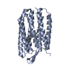
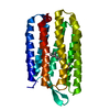
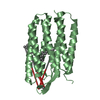
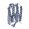
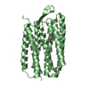
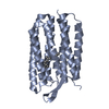
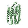
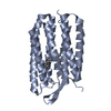
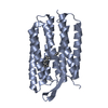
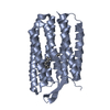
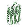
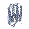
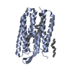
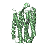
| Title | Crystal structure of bacteriorhodopsin mutant P186A crystallized from bicelles | ||||||
 Components Components | Bacteriorhodopsin | ||||||
 Keywords Keywords | MEMBRANE PROTEIN / alpha helix | ||||||
| Function / homology |  Function and homology informationphotoreceptor activity / phototransduction / proton transmembrane transport / monoatomic ion channel activity / plasma membrane Function and homology informationphotoreceptor activity / phototransduction / proton transmembrane transport / monoatomic ion channel activity / plasma membraneSimilarity search - Function | ||||||
| Biological species |  Halobacterium salinarum (Halophile) Halobacterium salinarum (Halophile) | ||||||
| Method | X-RAY DIFFRACTION / SYNCHROTRON / MOLECULAR REPLACEMENT / Resolution: 2.3 Å | ||||||
 Authors Authors | Yohannan, S. / Faham, S. / Yang, D. / Whitelegge, J.P. / Bowie, J.U. | ||||||
 Citation Citation | Journal: Proc.Natl.Acad.Sci.USA / Year: 2004 Title: The evolution of transmembrane helix kinks and the structural diversity of G protein-coupled receptors. Authors: Yohannan, S. / Faham, S. / Yang, D. / Whitelegge, J.P. / Bowie, J.U. | ||||||
| History |
|
- Structure visualization
| Structure viewer | Molecule: MolmilJmol/JSmol |
|---|
- Downloads & links
-Download
| PDBx/mmCIF format | 1q5i.cif.gz | 98.7 KB | Display | PDBx/mmCIF format |
|---|---|---|---|---|
| PDB format | pdb1q5i.ent.gz | 76.4 KB | Display | PDB format |
| PDBx/mmJSON format | 1q5i.json.gz | Tree view | PDBx/mmJSON format | |
| Others |  Other downloads Other downloads |
-Validation report
| Arichive directory | https://data.pdbj.org/pub/pdb/validation_reports/q5/1q5iftp://data.pdbj.org/pub/pdb/validation_reports/q5/1q5i | HTTPS FTP |
|---|
-Related structure data
| Related structure data | 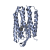 1q5jC  1py6S C: citing same article ( S: Starting model for refinement |
|---|---|
| Similar structure data |


-Links
 PDBj
PDBj


- Assembly
| Deposited unit | 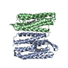
| ||||||||
|---|---|---|---|---|---|---|---|---|---|
| 1 | 
| ||||||||
| 2 | 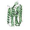
| ||||||||
| Unit cell |
|
-Components
| #1: Protein | / BR Mass: 26903.463 Da / Num. of mol.: 2 / Mutation: P186A Source method: isolated from a genetically manipulated source Source: (gene. exp.) Halobacterium salinarum (Halophile) / Strain: L33 / Gene: BOP OR VNG1467G / Plasmid: pMPK85 / Production host:  Escherichia coli (E. coli) / References: UniProt: P02945 Escherichia coli (E. coli) / References: UniProt: P02945#2: Chemical | Retinal  Mass: 284.436 Da / Num. of mol.: 2 / Source method: obtained synthetically / Formula: C20H28O Mass: 284.436 Da / Num. of mol.: 2 / Source method: obtained synthetically / Formula: C20H28O#3: Water | ChemComp-HOH / | Water Mass: 18.015 Da / Num. of mol.: 84 / Source method: isolated from a natural source / Formula: H2O Mass: 18.015 Da / Num. of mol.: 84 / Source method: isolated from a natural source / Formula: H2O |
|---|
-Experimental details
-Experiment
| Experiment | Method: X-RAY DIFFRACTION / Number of used crystals: 1 |
|---|
- Sample preparation
| Crystal | Density Matthews: 2.32 Å3/Da / Density % sol: 46.96 % | ||||||||||||||||||||||||||||
|---|---|---|---|---|---|---|---|---|---|---|---|---|---|---|---|---|---|---|---|---|---|---|---|---|---|---|---|---|---|
| Crystal grow | Temperature: 310 K / Method: vapor diffusion, hanging drop / pH: 4 Details: DMPC, CHAPSO, sodium phosphate, hexanediol, pH 4.0, VAPOR DIFFUSION, HANGING DROP, temperature 310.0K | ||||||||||||||||||||||||||||
| Crystal grow | *PLUS pH: 3.7 / Method: unknown | ||||||||||||||||||||||||||||
| Components of the solutions | *PLUS
|
-Data collection
| Diffraction | Mean temperature: 100 K |
|---|---|
| Diffraction source | Source: SYNCHROTRON / Site: ALS  / Beamline: 8.2.2 / Wavelength: 1.0332 Å / Beamline: 8.2.2 / Wavelength: 1.0332 Å |
| Detector | Type: ADSC QUANTUM 315 / Detector: CCD / Date: May 22, 2003 |
| Radiation | Monochromator: Double-crystal Si(111) / Protocol: SINGLE WAVELENGTH / Monochromatic (M) / Laue (L): M / Scattering type: x-ray |
| Radiation wavelength | Wavelength: 1.0332 Å / Relative weight: 1 |
| Reflection | Resolution: 2.2→500 Å / Num. obs: 24112 / % possible obs: 96.6 % / Observed criterion σ(F): 0 / Observed criterion σ(I): 0 / Redundancy: 4 % / Rmerge(I) obs: 0.116 / Net I/σ(I): 23 |
| Reflection shell | Resolution: 2.2→2.28 Å / % possible all: 92.4 |
- Processing
| Software |
| ||||||||||||||||||||||||||||||||||||
|---|---|---|---|---|---|---|---|---|---|---|---|---|---|---|---|---|---|---|---|---|---|---|---|---|---|---|---|---|---|---|---|---|---|---|---|---|---|
| Refinement | Method to determine structure: MOLECULAR REPLACEMENT Starting model: PDB ENTRY 1PY6 Resolution: 2.3→500 Å / Cross valid method: THROUGHOUT / σ(F): 0 / Stereochemistry target values: Engh & Huber Details: Crystal twinning was observed. Twinning operation = -h, -k, h+l. Twinning fraction = 0.5
| ||||||||||||||||||||||||||||||||||||
| Refine analyze |
| ||||||||||||||||||||||||||||||||||||
| Refinement step | Cycle: LAST / Resolution: 2.3→500 Å
| ||||||||||||||||||||||||||||||||||||
| Refine LS restraints |
| ||||||||||||||||||||||||||||||||||||
| LS refinement shell | Highest resolution: 2.3 Å
| ||||||||||||||||||||||||||||||||||||
| Xplor file |
| ||||||||||||||||||||||||||||||||||||
| Refinement | *PLUS Rfactor Rfree: 0.255 / Rfactor Rwork: 0.197 | ||||||||||||||||||||||||||||||||||||
| Solvent computation | *PLUS | ||||||||||||||||||||||||||||||||||||
| Displacement parameters | *PLUS | ||||||||||||||||||||||||||||||||||||
| Refine LS restraints | *PLUS
|
