 Movie
Movie Controller

[English] 日本語
 Yorodumi
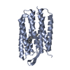
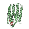
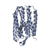
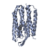
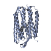
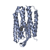
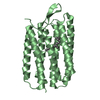
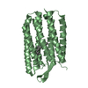
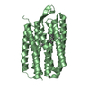
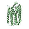
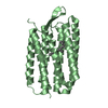
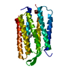
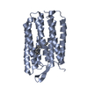
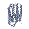
- PDB-3han: Crystal structure of bacteriorhodopsin mutant V49A crystallized f... -
+ Open data

- Basic information
| Entry | Database: PDB / ID: 3han | ||||||
|---|---|---|---|---|---|---|---|
| Title | Crystal structure of bacteriorhodopsin mutant V49A crystallized from bicelles | ||||||
 Components Components | Bacteriorhodopsin | ||||||
 Keywords Keywords | TRANSPORT PROTEIN / bacteriorhodopsin / packing force / van der Waals / evolutionary constraint / membrane protein / integral membrane protein / helical membrane protein / proton transport / Cell membrane / Chromophore / Hydrogen ion transport / Ion transport / Membrane / Photoreceptor protein / Pyrrolidone carboxylic acid / Receptor / Retinal protein / Sensory transduction / Transmembrane / Transport | ||||||
| Function / homology |  Function and homology informationphotoreceptor activity / phototransduction / proton transmembrane transport / monoatomic ion channel activity / plasma membrane Function and homology informationphotoreceptor activity / phototransduction / proton transmembrane transport / monoatomic ion channel activity / plasma membraneSimilarity search - Function | ||||||
| Biological species |  Halobacterium salinarum (Halophile) Halobacterium salinarum (Halophile) | ||||||
| Method | X-RAY DIFFRACTION / SYNCHROTRON / MOLECULAR REPLACEMENT / Resolution: 2.75 Å | ||||||
 Authors Authors | Joh, N.H. / Yang, D. / Bowie, J.U. | ||||||
 Citation Citation | Journal: J.Am.Chem.Soc. / Year: 2009 Title: Similar energetic contributions of packing in the core of membrane and water-soluble proteins. Authors: Joh, N.H. / Oberai, A. / Yang, D. / Whitelegge, J.P. / Bowie, J.U. | ||||||
| History |
|
- Structure visualization
| Structure viewer | Molecule: MolmilJmol/JSmol |
|---|
- Downloads & links
-Download
| PDBx/mmCIF format | 3han.cif.gz | 53 KB | Display | PDBx/mmCIF format |
|---|---|---|---|---|
| PDB format | pdb3han.ent.gz | 41.3 KB | Display | PDB format |
| PDBx/mmJSON format | 3han.json.gz | Tree view | PDBx/mmJSON format | |
| Others |  Other downloads Other downloads |
-Validation report
| Arichive directory | https://data.pdbj.org/pub/pdb/validation_reports/ha/3hanftp://data.pdbj.org/pub/pdb/validation_reports/ha/3han | HTTPS FTP |
|---|
-Related structure data


-Links
 PDBj
PDBj


- Assembly
| Deposited unit | 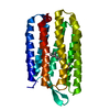
| ||||||||
|---|---|---|---|---|---|---|---|---|---|
| 1 |
| ||||||||
| Unit cell |
|
-Components
| #1: Protein | / BR Mass: 26901.445 Da / Num. of mol.: 1 / Mutation: V49A Source method: isolated from a genetically manipulated source Source: (gene. exp.) Halobacterium salinarum (Halophile) / Gene: bop, VNG_1467G / Production host: Halobacterium salinarum (Halophile) / Strain (production host): L33 / References: UniProt: P02945 |
|---|---|
| #2: Chemical | ChemComp-RET / Retinal  Mass: 284.436 Da / Num. of mol.: 1 / Source method: obtained synthetically / Formula: C20H28O Mass: 284.436 Da / Num. of mol.: 1 / Source method: obtained synthetically / Formula: C20H28O |
| #3: Water | ChemComp-HOH / Water Mass: 18.015 Da / Num. of mol.: 8 / Source method: isolated from a natural source / Formula: H2O Mass: 18.015 Da / Num. of mol.: 8 / Source method: isolated from a natural source / Formula: H2O |
-Experimental details
-Experiment
| Experiment | Method: X-RAY DIFFRACTION / Number of used crystals: 1 |
|---|
- Sample preparation
| Crystal | Density Matthews: 2.76 Å3/Da / Density % sol: 55.42 % |
|---|---|
| Crystal grow | Temperature: 310 K / Method: hanging drop, bicelle method Details: 1 M sodium phosphate (pH 3.9), 1 M sodium phosphate (pH 4.5), 180 mM 1,6-hexanediol, 3.5% triethyleneglycol, PFPC used as cryoprotectant, hanging drop, bicelle method, temperature 310K |
-Data collection
| Diffraction | Mean temperature: 100 K |
|---|---|
| Diffraction source | Source: SYNCHROTRON / Site: APS  / Beamline: 24-ID-C / Wavelength: 0.9793 Å / Beamline: 24-ID-C / Wavelength: 0.9793 Å |
| Detector | Type: ADSC QUANTUM 315 / Detector: CCD / Date: Jun 25, 2008 |
| Radiation | Monochromator: Si(111) double crystal / Protocol: SINGLE WAVELENGTH / Monochromatic (M) / Laue (L): M / Scattering type: x-ray |
| Radiation wavelength | Wavelength: 0.9793 Å / Relative weight: 1 |
| Reflection | Resolution: 2.75→50 Å / Num. obs: 7285 / % possible obs: 89.9 % / Redundancy: 5.8 % / Rmerge(I) obs: 0.105 / Χ2: 1.023 / Net I/σ(I): 10.238 |
| Reflection shell | Resolution: 2.75→2.85 Å / Redundancy: 3.1 % / Rmerge(I) obs: 0.382 / Num. unique all: 421 / Χ2: 0.932 / % possible all: 52.8 |
- Processing
| Software |
| |||||||||||||||||||||||||||||||||||||||||||||||||||||||||||||||||||||||||||||||||||||
|---|---|---|---|---|---|---|---|---|---|---|---|---|---|---|---|---|---|---|---|---|---|---|---|---|---|---|---|---|---|---|---|---|---|---|---|---|---|---|---|---|---|---|---|---|---|---|---|---|---|---|---|---|---|---|---|---|---|---|---|---|---|---|---|---|---|---|---|---|---|---|---|---|---|---|---|---|---|---|---|---|---|---|---|---|---|---|
| Refinement | Method to determine structure: MOLECULAR REPLACEMENT / Resolution: 2.75→41.2 Å / Cor.coef. Fo:Fc: 0.919 / Cor.coef. Fo:Fc free: 0.879 / Occupancy max: 1 / Occupancy min: 1 / SU B: 13.038 / SU ML: 0.276 / Cross valid method: THROUGHOUT / σ(F): 0 / ESU R Free: 0.426 Stereochemistry target values: MAXIMUM LIKELIHOOD WITH PHASES Details: HYDROGENS HAVE BEEN ADDED IN THE RIDING POSITIONS
| |||||||||||||||||||||||||||||||||||||||||||||||||||||||||||||||||||||||||||||||||||||
| Solvent computation | Ion probe radii: 0.8 Å / Shrinkage radii: 0.8 Å / VDW probe radii: 1.4 Å / Solvent model: MASK | |||||||||||||||||||||||||||||||||||||||||||||||||||||||||||||||||||||||||||||||||||||
| Displacement parameters | Biso max: 88.96 Å2 / Biso mean: 40.424 Å2 / Biso min: 18.3 Å2
| |||||||||||||||||||||||||||||||||||||||||||||||||||||||||||||||||||||||||||||||||||||
| Refinement step | Cycle: LAST / Resolution: 2.75→41.2 Å
| |||||||||||||||||||||||||||||||||||||||||||||||||||||||||||||||||||||||||||||||||||||
| Refine LS restraints |
| |||||||||||||||||||||||||||||||||||||||||||||||||||||||||||||||||||||||||||||||||||||
| LS refinement shell | Resolution: 2.75→2.816 Å / Total num. of bins used: 20
|
