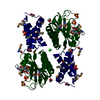
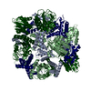
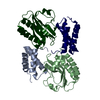
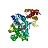
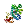
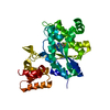
- PDB-6psh: Crystal structure of periplasmic domain of antiholin RI from T4 phage -
+
データを開く
IDまたはキーワード:
読み込み中...
-
基本情報
登録情報
データベース: PDB / ID: 6psh
タイトル
Crystal structure of periplasmic domain of antiholin RI from T4 phage
要素
Antiholin
キーワード
VIRAL PROTEIN (ウイルスタンパク質) / phage (ファージ) / lysis inhibition
機能・相同性
Antiholin T4 type / host cell periplasmic space / molecular function inhibitor activity / killing of cells of another organism / host cell plasma membrane / DNA binding / Antiholin
ジャーナル: J Mol Biol / 年: 2020 タイトル: The Structural Basis of T4 Phage Lysis Control: DNA as the Signal for Lysis Inhibition. 著者: Inna V Krieger / Vladimir Kuznetsov / Jeng-Yih Chang / Junjie Zhang / Samir H Moussa / Ryland F Young / James C Sacchettini / 要旨: Optimal phage propagation depends on the regulation of the lysis of the infected host cell. In T4 phage infection, lysis occurs when the holin protein (T) forms lesions in the host membrane. However, ...Optimal phage propagation depends on the regulation of the lysis of the infected host cell. In T4 phage infection, lysis occurs when the holin protein (T) forms lesions in the host membrane. However, the lethal function of T can be blocked by an antiholin (RI) during lysis inhibition (LIN). LIN sets if the infected cell undergoes superinfection, then the lysis is delayed until host/phage ratio becomes more favorable for the release of progeny. It has been thought that a signal derived from the superinfection is required to activate RI. Here we report structures that suggest a radically different model in which RI binds to T irrespective of superinfection, causing it to accumulate in a membrane as heterotetrameric 2RI-2T complex. Moreover, we show the complex binds non-specifically to DNA, suggesting that the gDNA from the superinfecting phage serves as the LIN signal and that stabilization of the complex by DNA binding is what defines LIN. Finally, we show that soluble domain of free RI crystallizes in a domain-swapped homotetramer, which likely works as a sink for RI molecules released from the RI-T complex to ensure efficient lysis. These results constitute the first structural basis and a new model not only for the historic LIN phenomenon but also for the temporal regulation of phage lysis in general.
 ムービー
ムービー コントローラー


 データを開く
 基本情報
基本情報 要素
要素 キーワード
キーワード VIRAL PROTEIN (ウイルスタンパク質) /
  機能・相同性情報
 データ登録者
 米国, 1件
  引用
引用 構造の表示
構造の表示 ダウンロードとリンク
 その他のダウンロード
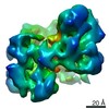


 PDBj
PDBj 集合体
集合体


 分子量: 62.068 Da / 分子数: 1 / 由来タイプ: 天然 / 式: C2H6O2
 分子量: 18.015 Da / 分子数: 10 / 由来タイプ: 天然 / 式: H2O
 試料調製
試料調製 解析
解析