 Movie
Movie Controller

[English] 日本語
 Yorodumi
- PDB-6pjq: Time-resolved structural snapshot of proteolysis by GlpG inside t... -
+ Open data

- Basic information
| Entry | Database: PDB / ID: 6pjq | ||||||
|---|---|---|---|---|---|---|---|
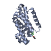
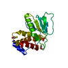
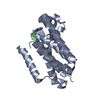
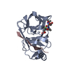
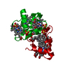
| Title | Time-resolved structural snapshot of proteolysis by GlpG inside the membrane | ||||||
 Components Components |
| ||||||
 Keywords Keywords |  MEMBRANE PROTEIN / substrate complex MEMBRANE PROTEIN / substrate complex | ||||||
| Function / homology |  Function and homology information Function and homology informationmaternal determination of dorsal/ventral axis, ovarian follicular epithelium, germ-line encoded / dorsal/ventral axis specification, ovarian follicular epithelium / anterior/posterior axis specification, follicular epithelium / determination of dorsal identity / chorion-containing eggshell pattern formation / oocyte anterior/posterior axis specification / oocyte microtubule cytoskeleton organization / oocyte dorsal/ventral axis specification / imaginal disc-derived wing vein specification / dorsal appendage formation ...maternal determination of dorsal/ventral axis, ovarian follicular epithelium, germ-line encoded / dorsal/ventral axis specification, ovarian follicular epithelium / anterior/posterior axis specification, follicular epithelium / determination of dorsal identity / chorion-containing eggshell pattern formation / oocyte anterior/posterior axis specification / oocyte microtubule cytoskeleton organization / oocyte dorsal/ventral axis specification / imaginal disc-derived wing vein specification / dorsal appendage formation / positive regulation of border follicle cell migration / rhomboid protease / anterior/posterior pattern specification / epidermal growth factor receptor binding / epidermal growth factor receptor signaling pathway / endopeptidase activity / receptor ligand activity / cell surface receptor signaling pathway / serine-type endopeptidase activity / proteolysis / extracellular space / identical protein binding / plasma membraneSimilarity search - Function | ||||||
| Biological species |  Escherichia coli (E. coli) Escherichia coli (E. coli) Drosophila melanogaster (fruit fly) Drosophila melanogaster (fruit fly) | ||||||
| Method | X-RAY DIFFRACTION / SYNCHROTRON / MOLECULAR REPLACEMENT / Resolution: 2.5 Å | ||||||
 Authors Authors | Urban, S. / Cho, S. | ||||||
| Funding support |  United States, 1items United States, 1items
| ||||||
 Citation Citation | Journal: Nat.Struct.Mol.Biol. / Year: 2019 Title: Ten catalytic snapshots of rhomboid intramembrane proteolysis from gate opening to peptide release. Authors: Cho, S. / Baker, R.P. / Ji, M. / Urban, S. | ||||||
| History |
|
- Structure visualization
| Structure viewer | Molecule: MolmilJmol/JSmol |
|---|
- Downloads & links
-Download
| PDBx/mmCIF format | 6pjq.cif.gz | 51.5 KB | Display | PDBx/mmCIF format |
|---|---|---|---|---|
| PDB format | pdb6pjq.ent.gz | 34.2 KB | Display | PDB format |
| PDBx/mmJSON format | 6pjq.json.gz | Tree view | PDBx/mmJSON format | |
| Others |  Other downloads Other downloads |
-Validation report
| Arichive directory | https://data.pdbj.org/pub/pdb/validation_reports/pj/6pjqftp://data.pdbj.org/pub/pdb/validation_reports/pj/6pjq | HTTPS FTP |
|---|
-Related structure data
| Related structure data | 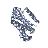 6pj4C  6pj5C 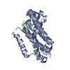 6pj7C 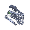 6pj8C  6pj9C 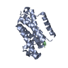 6pjaC 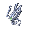 6pjpC 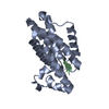 6pjrC 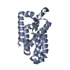 6pjuC 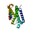 5f5dS S: Starting model for refinement C: citing same article ( |
|---|---|
| Similar structure data |


-Links
 PDBj
PDBj
- Assembly
| Deposited unit | 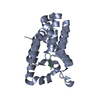
| ||||||||
|---|---|---|---|---|---|---|---|---|---|
| 1 |
| ||||||||
| Unit cell |
|
-Components
| #1: Protein | Mass: 23800.133 Da / Num. of mol.: 1 Source method: isolated from a genetically manipulated source Source: (gene. exp.) Escherichia coli (E. coli) / Gene: glpG, APT88_21985, SK83_00858 / Production host: Escherichia coli BL21(DE3) (bacteria) / Strain (production host): BL21(DE3)References: UniProt: A0A0J2E248, UniProt: P09391*PLUS, rhomboid protease |
|---|---|
| #2: Protein/peptide | Mass: 1524.894 Da / Num. of mol.: 1 / Source method: obtained synthetically / Source: (synth.) Drosophila melanogaster (fruit fly) / References: UniProt: P42287*PLUS |
| #3: Water | ChemComp-HOH / Water Mass: 18.015 Da / Num. of mol.: 16 / Source method: isolated from a natural source / Formula: H2O Mass: 18.015 Da / Num. of mol.: 16 / Source method: isolated from a natural source / Formula: H2O |
-Experimental details
-Experiment
| Experiment | Method: X-RAY DIFFRACTION / Number of used crystals: 1 |
|---|
- Sample preparation
| Crystal | Density Matthews: 2.14 Å3/Da / Density % sol: 42.44 % |
|---|---|
| Crystal grow | Temperature: 298 K / Method: vapor diffusion, hanging drop / pH: 5.5 Details: 0.1 M Na-acetate pH 5.5, 3 M NaCl, and 5 % ethylene glycol |
-Data collection
| Diffraction | Mean temperature: 100 K / Serial crystal experiment: N |
|---|---|
| Diffraction source | Source: SYNCHROTRON / Site: CHESS / Beamline: F1 / Wavelength: 0.977 Å |
| Detector | Type: DECTRIS PILATUS 6M / Detector: PIXEL / Date: May 21, 2018 |
| Radiation | Protocol: SINGLE WAVELENGTH / Monochromatic (M) / Laue (L): M / Scattering type: x-ray |
| Radiation wavelength | Wavelength: 0.977 Å / Relative weight: 1 |
| Reflection | Resolution: 2.5→57.63 Å / Num. obs: 7484 / % possible obs: 96 % / Redundancy: 5.6 % / Rsym value: 0.041 / Net I/σ(I): 10.3 |
| Reflection shell | Resolution: 2.5→2.6 Å / Num. unique obs: 851 / Rsym value: 0.451 |
- Processing
| Software |
| |||||||||||||||||||||||||||||||||||||||||||||
|---|---|---|---|---|---|---|---|---|---|---|---|---|---|---|---|---|---|---|---|---|---|---|---|---|---|---|---|---|---|---|---|---|---|---|---|---|---|---|---|---|---|---|---|---|---|---|
| Refinement | Method to determine structure: MOLECULAR REPLACEMENT Starting model: 5F5D Resolution: 2.5→57.56 Å / Cor.coef. Fo:Fc: 0.908 / Cor.coef. Fo:Fc free: 0.844 / SU B: 13.201 / SU ML: 0.271 / Cross valid method: THROUGHOUT / σ(F): 0 / ESU R: 0.576 / ESU R Free: 0.325 / Stereochemistry target values: MAXIMUM LIKELIHOOD / Details: U VALUES : REFINED INDIVIDUALLY
| |||||||||||||||||||||||||||||||||||||||||||||
| Solvent computation | Ion probe radii: 0.8 Å / Shrinkage radii: 0.8 Å / VDW probe radii: 1.2 Å / Solvent model: MASK | |||||||||||||||||||||||||||||||||||||||||||||
| Displacement parameters | Biso max: 162.57 Å2 / Biso mean: 69.509 Å2 / Biso min: 36.2 Å2
| |||||||||||||||||||||||||||||||||||||||||||||
| Refinement step | Cycle: final / Resolution: 2.5→57.56 Å
| |||||||||||||||||||||||||||||||||||||||||||||
| Refine LS restraints |
| |||||||||||||||||||||||||||||||||||||||||||||
| LS refinement shell | Resolution: 2.5→2.565 Å / Rfactor Rfree error: 0 / Total num. of bins used: 20
|
