 Movie
Movie Controller

[English] 日本語
 Yorodumi
- PDB-6vj9: Crystal structure of GlpG in complex with peptide boronate inhibitor -
+ Open data

- Basic information
| Entry | Database: PDB / ID: 6vj9 | ||||||
|---|---|---|---|---|---|---|---|
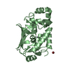
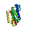
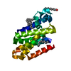
| Title | Crystal structure of GlpG in complex with peptide boronate inhibitor | ||||||
 Components Components |
| ||||||
 Keywords Keywords | HYDROLASE/HYDROLASE INHIBITOR /  inhibitor / complex / GlpG / rhomboid protease / HYDROLASE-HYDROLASE INHIBITOR complex inhibitor / complex / GlpG / rhomboid protease / HYDROLASE-HYDROLASE INHIBITOR complex | ||||||
| Function / homology |  Function and homology informationrhomboid protease / endopeptidase activity / membrane => GO:0016020 / serine-type endopeptidase activity / proteolysis / identical protein binding / plasma membrane Function and homology informationrhomboid protease / endopeptidase activity / membrane => GO:0016020 / serine-type endopeptidase activity / proteolysis / identical protein binding / plasma membraneSimilarity search - Function | ||||||
| Biological species |  Escherichia coli (E. coli) Escherichia coli (E. coli) Drosophila melanogaster (fruit fly) Drosophila melanogaster (fruit fly) | ||||||
| Method | X-RAY DIFFRACTION / SYNCHROTRON / MOLECULAR REPLACEMENT / Resolution: 2.3 Å | ||||||
 Authors Authors | Urban, S. / Cho, S. | ||||||
| Funding support |  United States, 1items United States, 1items
| ||||||
 Citation Citation | Journal: Cell Chem Biol / Year: 2020 Title: Designed Parasite-Selective Rhomboid Inhibitors Block Invasion and Clear Blood-Stage Malaria. Authors: Gandhi, S. / Baker, R.P. / Cho, S. / Stanchev, S. / Strisovsky, K. / Urban, S. | ||||||
| History |
|
- Structure visualization
| Structure viewer | Molecule: MolmilJmol/JSmol |
|---|
- Downloads & links
-Download
| PDBx/mmCIF format | 6vj9.cif.gz | 53.5 KB | Display | PDBx/mmCIF format |
|---|---|---|---|---|
| PDB format | pdb6vj9.ent.gz | 36.3 KB | Display | PDB format |
| PDBx/mmJSON format | 6vj9.json.gz | Tree view | PDBx/mmJSON format | |
| Others |  Other downloads Other downloads |
-Validation report
| Arichive directory | https://data.pdbj.org/pub/pdb/validation_reports/vj/6vj9ftp://data.pdbj.org/pub/pdb/validation_reports/vj/6vj9 | HTTPS FTP |
|---|
-Related structure data
| Related structure data |  6vj8C  6xroC  6xrpC  2ic8S S: Starting model for refinement C: citing same article ( |
|---|---|
| Similar structure data |


-Links
 PDBj
PDBj
- Assembly
| Deposited unit | 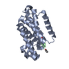
| ||||||||
|---|---|---|---|---|---|---|---|---|---|
| 1 |
| ||||||||
| Unit cell |
| ||||||||
| Components on special symmetry positions |
|
-Components
| #1: Protein | Mass: 21927.043 Da / Num. of mol.: 1 / Fragment: UNP residue 61-250 Source method: isolated from a genetically manipulated source Source: (gene. exp.) Escherichia coli (E. coli) / Gene: glpG, EVY14_07190 / Production host: Escherichia coli BL21(DE3) (bacteria) / Strain (production host): C43(DE3) / References: UniProt: A0A4Q6HQV3, UniProt: P09391*PLUS |
|---|---|
| #2: Protein/peptide |   Type: Oligopeptide / Class: Inhibitor / Mass: 502.459 Da / Num. of mol.: 1 / Source method: obtained synthetically / Source: (synth.) Drosophila melanogaster (fruit fly) / References: ACE-VAL-ARG-MET-B2A Type: Oligopeptide / Class: Inhibitor / Mass: 502.459 Da / Num. of mol.: 1 / Source method: obtained synthetically / Source: (synth.) Drosophila melanogaster (fruit fly) / References: ACE-VAL-ARG-MET-B2A |
| #3: Water | ChemComp-HOH / Water Mass: 18.015 Da / Num. of mol.: 19 / Source method: isolated from a natural source / Formula: H2O Mass: 18.015 Da / Num. of mol.: 19 / Source method: isolated from a natural source / Formula: H2O |
| Has ligand of interest | Y |
-Experimental details
-Experiment
| Experiment | Method: X-RAY DIFFRACTION / Number of used crystals: 1 |
|---|
- Sample preparation
| Crystal | Density Matthews: 3.33 Å3/Da / Density % sol: 63.04 % |
|---|---|
| Crystal grow | Temperature: 298 K / Method: vapor diffusion, hanging drop Details: 0.1 M Tris, pH 8.5, 3 M sodium nitrate, 15% glycerol |
-Data collection
| Diffraction | Mean temperature: 100 K / Serial crystal experiment: N |
|---|---|
| Diffraction source | Source: SYNCHROTRON / Site: CHESS / Beamline: F1 / Wavelength: 0.9718 Å |
| Detector | Type: DECTRIS PILATUS 6M / Detector: PIXEL / Date: May 7, 2018 |
| Radiation | Monochromator: Si(111) / Protocol: SINGLE WAVELENGTH / Monochromatic (M) / Laue (L): M / Scattering type: x-ray |
| Radiation wavelength | Wavelength: 0.9718 Å / Relative weight: 1 |
| Reflection | Resolution: 2.3→32.13 Å / Num. obs: 13444 / % possible obs: 99.9 % / Redundancy: 8 % / Rmerge(I) obs: 0.093 / Net I/σ(I): 7.5 |
| Reflection shell | Resolution: 2.3→2.38 Å / Rmerge(I) obs: 0.621 / Num. unique obs: 1296 |
- Processing
| Software |
| ||||||||||||||||||||||||||||||||||||||||||||||||||||||||||||||||||||||||||||||||||||||||||||||||||||||||||||||||||||||||||||||||||||||||||||||||||||||||||||||||||||||||||||||||||||||
|---|---|---|---|---|---|---|---|---|---|---|---|---|---|---|---|---|---|---|---|---|---|---|---|---|---|---|---|---|---|---|---|---|---|---|---|---|---|---|---|---|---|---|---|---|---|---|---|---|---|---|---|---|---|---|---|---|---|---|---|---|---|---|---|---|---|---|---|---|---|---|---|---|---|---|---|---|---|---|---|---|---|---|---|---|---|---|---|---|---|---|---|---|---|---|---|---|---|---|---|---|---|---|---|---|---|---|---|---|---|---|---|---|---|---|---|---|---|---|---|---|---|---|---|---|---|---|---|---|---|---|---|---|---|---|---|---|---|---|---|---|---|---|---|---|---|---|---|---|---|---|---|---|---|---|---|---|---|---|---|---|---|---|---|---|---|---|---|---|---|---|---|---|---|---|---|---|---|---|---|---|---|---|---|
| Refinement | Method to determine structure: MOLECULAR REPLACEMENT Starting model: PDB entry 2IC8 Resolution: 2.3→32.15 Å / Cor.coef. Fo:Fc: 0.934 / Cor.coef. Fo:Fc free: 0.933 / SU B: 10.514 / SU ML: 0.227 / Cross valid method: THROUGHOUT / ESU R: 0.247 / ESU R Free: 0.206
| ||||||||||||||||||||||||||||||||||||||||||||||||||||||||||||||||||||||||||||||||||||||||||||||||||||||||||||||||||||||||||||||||||||||||||||||||||||||||||||||||||||||||||||||||||||||
| Solvent computation | Ion probe radii: 0.8 Å / Shrinkage radii: 0.8 Å / VDW probe radii: 1.2 Å | ||||||||||||||||||||||||||||||||||||||||||||||||||||||||||||||||||||||||||||||||||||||||||||||||||||||||||||||||||||||||||||||||||||||||||||||||||||||||||||||||||||||||||||||||||||||
| Displacement parameters | Biso mean: 62.98 Å2
| ||||||||||||||||||||||||||||||||||||||||||||||||||||||||||||||||||||||||||||||||||||||||||||||||||||||||||||||||||||||||||||||||||||||||||||||||||||||||||||||||||||||||||||||||||||||
| Refinement step | Cycle: 1 / Resolution: 2.3→32.15 Å
| ||||||||||||||||||||||||||||||||||||||||||||||||||||||||||||||||||||||||||||||||||||||||||||||||||||||||||||||||||||||||||||||||||||||||||||||||||||||||||||||||||||||||||||||||||||||
| Refine LS restraints |
|