 Movie
Movie Controller


+ Open data

- Basic information
| Entry | Database: PDB / ID: 6omi | ||||||
|---|---|---|---|---|---|---|---|
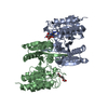
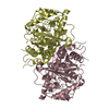
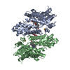
| Title | Crystal structure of the Legionella effector protein MavL | ||||||
 Components Components | MavL | ||||||
 Keywords Keywords |  HYDROLASE / ADP-ribosyltransferase (ART) / macrodomain / Secreted bacterial effector protein HYDROLASE / ADP-ribosyltransferase (ART) / macrodomain / Secreted bacterial effector protein | ||||||
| Function / homology | BROMIDE ION / Uncharacterized protein Function and homology information Function and homology information | ||||||
| Biological species |  Legionella pneumophila subsp. pneumophila (bacteria) Legionella pneumophila subsp. pneumophila (bacteria) | ||||||
| Method | X-RAY DIFFRACTION / SYNCHROTRON / SAD / Resolution: 2.643 Å | ||||||
 Authors Authors | Cygler, M. / Voth, K. | ||||||
| Funding support |  Canada, 1items Canada, 1items
| ||||||
 Citation Citation | Journal: To Be Published Title: Crystal Structure of the Legionella Effector protein MavL Authors: Cygler, M. / Voth, K. | ||||||
| History |
|
- Structure visualization
| Structure viewer | Molecule: MolmilJmol/JSmol |
|---|
- Downloads & links
-Download
| PDBx/mmCIF format | 6omi.cif.gz | 422.8 KB | Display | PDBx/mmCIF format |
|---|---|---|---|---|
| PDB format | pdb6omi.ent.gz | 361.6 KB | Display | PDB format |
| PDBx/mmJSON format | 6omi.json.gz | Tree view | PDBx/mmJSON format | |
| Others |  Other downloads Other downloads |
-Validation report
| Arichive directory | https://data.pdbj.org/pub/pdb/validation_reports/om/6omiftp://data.pdbj.org/pub/pdb/validation_reports/om/6omi | HTTPS FTP |
|---|
-Related structure data
| Similar structure data |
|---|


-Links
 PDBj
PDBj

- Assembly
| Deposited unit | 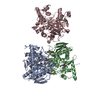
| |||||||||
|---|---|---|---|---|---|---|---|---|---|---|
| 1 | 
| |||||||||
| 2 | 
| |||||||||
| Unit cell |
| |||||||||
| Components on special symmetry positions |
|
-Components
| #1: Protein | Mass: 44423.336 Da / Num. of mol.: 3 / Fragment: residues 42-435 Source method: isolated from a genetically manipulated source Source: (gene. exp.) Legionella pneumophila subsp. pneumophila (strain Philadelphia 1 / ATCC 33152 / DSM 7513) (bacteria)Strain: Philadelphia 1 / ATCC 33152 / DSM 7513 / Gene: lpg2526 / Production host: Escherichia coli BL21(DE3) (bacteria) / Strain (production host): BL21(DE3) / References: UniProt: Q5ZSJ1#2: Chemical | Bromide  Mass: 79.904 Da / Num. of mol.: 2 / Source method: obtained synthetically / Formula: Br Mass: 79.904 Da / Num. of mol.: 2 / Source method: obtained synthetically / Formula: Br#3: Water | ChemComp-HOH / | Water Mass: 18.015 Da / Num. of mol.: 14 / Source method: isolated from a natural source / Formula: H2O Mass: 18.015 Da / Num. of mol.: 14 / Source method: isolated from a natural source / Formula: H2OHas ligand of interest | N | |
|---|
-Experimental details
-Experiment
| Experiment | Method: X-RAY DIFFRACTION / Number of used crystals: 1 |
|---|
- Sample preparation
| Crystal | Density Matthews: 3.02 Å3/Da / Density % sol: 59.23 % |
|---|---|
| Crystal grow | Temperature: 293.15 K / Method: vapor diffusion, hanging drop / pH: 6.5 Details: 20% PEG8000, 0.2M Ammonium sulfate, 0.1M MES pH 6.5 |
-Data collection
| Diffraction | Mean temperature: 293.15 K / Serial crystal experiment: N |
|---|---|
| Diffraction source | Source: SYNCHROTRON / Site: CLSI / Beamline: 08ID-1 / Wavelength: 0.993054 Å |
| Detector | Type: DECTRIS PILATUS3 S 6M / Detector: PIXEL / Date: Jan 15, 2018 |
| Radiation | Protocol: SINGLE WAVELENGTH / Monochromatic (M) / Laue (L): M / Scattering type: x-ray |
| Radiation wavelength | Wavelength: 0.993054 Å / Relative weight: 1 |
| Reflection | Resolution: 2.643→48.77 Å / Num. obs: 41740 / % possible obs: 99.26 % / Redundancy: 1.998 % / Rmerge(I) obs: 0.078 / Net I/σ(I): 21.53 |
| Reflection shell | Resolution: 2.643→2.738 Å / Rmerge(I) obs: 1.387 / Num. unique obs: 4058 |
- Processing
| Software |
| ||||||||||||||||||||||||||||||||||||||||||||||||||||||||||||||||||||||||||||||||||||||||||||||||||||
|---|---|---|---|---|---|---|---|---|---|---|---|---|---|---|---|---|---|---|---|---|---|---|---|---|---|---|---|---|---|---|---|---|---|---|---|---|---|---|---|---|---|---|---|---|---|---|---|---|---|---|---|---|---|---|---|---|---|---|---|---|---|---|---|---|---|---|---|---|---|---|---|---|---|---|---|---|---|---|---|---|---|---|---|---|---|---|---|---|---|---|---|---|---|---|---|---|---|---|---|---|---|
| Refinement | Method to determine structure: SAD / Resolution: 2.643→45.14 Å / SU ML: 0.41 / Cross valid method: FREE R-VALUE / σ(F): 1.35 / Phase error: 33.77 / Stereochemistry target values: ML
| ||||||||||||||||||||||||||||||||||||||||||||||||||||||||||||||||||||||||||||||||||||||||||||||||||||
| Solvent computation | Shrinkage radii: 0.9 Å / VDW probe radii: 1.11 Å / Solvent model: FLAT BULK SOLVENT MODEL | ||||||||||||||||||||||||||||||||||||||||||||||||||||||||||||||||||||||||||||||||||||||||||||||||||||
| Refinement step | Cycle: LAST / Resolution: 2.643→48.77 Å
| ||||||||||||||||||||||||||||||||||||||||||||||||||||||||||||||||||||||||||||||||||||||||||||||||||||
| Refine LS restraints |
| ||||||||||||||||||||||||||||||||||||||||||||||||||||||||||||||||||||||||||||||||||||||||||||||||||||
| LS refinement shell |
| ||||||||||||||||||||||||||||||||||||||||||||||||||||||||||||||||||||||||||||||||||||||||||||||||||||
| Refinement TLS params. | Method: refined / Refine-ID: X-RAY DIFFRACTION
| ||||||||||||||||||||||||||||||||||||||||||||||||||||||||||||||||||||||||||||||||||||||||||||||||||||
| Refinement TLS group |
|
