 Movie
Movie Controller

[English] 日本語
 Yorodumi
- PDB-2po7: Crystal structure of human ferrochelatase mutant with His 341 rep... -
+ Open data

- Basic information
| Entry | Database: PDB / ID: 2po7 | ||||||
|---|---|---|---|---|---|---|---|
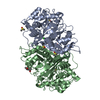
| Title | Crystal structure of human ferrochelatase mutant with His 341 replaced by Cys | ||||||
 Components Components | Ferrochelatase, mitochondrial | ||||||
 Keywords Keywords | LYASE / FERROCHELATASE / H341C / FE2S2 CLUSTER / HEME BIOSYNTHESIS / PROTOHEME / FERRO-LYASE / MATURE LENGTH / PROTEOLYTICALLY PROCESSED MITOCHONDRIAL INNER MEMBRANE PROTEIN | ||||||
| Function / homology |  Function and homology information Function and homology informationprotoporphyrinogen IX metabolic process / regulation of hemoglobin biosynthetic process / regulation of eIF2 alpha phosphorylation by heme / detection of UV / iron-responsive element binding / response to platinum ion / protoporphyrin ferrochelatase / response to insecticide / heme B biosynthetic process / heme O biosynthetic process ...protoporphyrinogen IX metabolic process / regulation of hemoglobin biosynthetic process / regulation of eIF2 alpha phosphorylation by heme / detection of UV / iron-responsive element binding / response to platinum ion / protoporphyrin ferrochelatase / response to insecticide / heme B biosynthetic process / heme O biosynthetic process / ferrochelatase activity / heme A biosynthetic process / very-low-density lipoprotein particle assembly / response to methylmercury / response to arsenic-containing substance / Heme biosynthesis / heme biosynthetic process / response to light stimulus / cholesterol metabolic process / erythrocyte differentiation / cellular response to dexamethasone stimulus / generation of precursor metabolites and energy / response to lead ion / ferrous iron binding / 2 iron, 2 sulfur cluster binding / multicellular organismal-level iron ion homeostasis / response to ethanol / mitochondrial inner membrane / mitochondrial matrix / response to xenobiotic stimulus / heme binding / protein homodimerization activity / mitochondrion / identical protein bindingSimilarity search - Function | ||||||
| Biological species |  Homo sapiens (human) Homo sapiens (human) | ||||||
| Method | X-RAY DIFFRACTION / DIFFERENCE FOURIER / Resolution: 2.2 Å | ||||||
 Authors Authors | Dailey, H.A. / Wu, C.-K. / Horanyi, P. / Medlock, A.E. / Najahi-Missaoui, W. / Burden, A. / Dailey, T.A. / Rose, J.P. | ||||||
 Citation Citation | Journal: Biochemistry / Year: 2007 Title: Altered orientation of active site residues in variants of human ferrochelatase. Evidence for a hydrogen bond network involved in catalysis. Authors: Dailey, H.A. / Wu, C.K. / Horanyi, P. / Medlock, A.E. / Najahi-Missaoui, W. / Burden, A.E. / Dailey, T.A. / Rose, J. #1: Journal: Nat.Struct.Biol. / Year: 2001Title: The 2.0 A structure of human ferrochelatase, the terminal enzyme of heme biosynthesis Authors: Wu, C.K. / Dailey, H.A. / Rose, J.P. / Burden, A. / Sellers, V.M. / Wang, B.-C. #2: Journal: Biochim.Biophys.Acta / Year: 1999 Title: Human Ferrochelatase: Crystallization, Characterization of the [2Fe-2S] Cluster and Determination that the Enzyme is a Homodimer Authors: Burden, A.E. / Wu, C.-K. / Dailey, T.A. / Busch, J.L.H. / Dhawan, I.K. / Rose, J.P. / Wang, B.C. / Dailey, H.A. | ||||||
| History |
|
- Structure visualization
| Structure viewer | Molecule: MolmilJmol/JSmol |
|---|
- Downloads & links
-Download
| PDBx/mmCIF format | 2po7.cif.gz | 167.2 KB | Display | PDBx/mmCIF format |
|---|---|---|---|---|
| PDB format | pdb2po7.ent.gz | 130.3 KB | Display | PDB format |
| PDBx/mmJSON format | 2po7.json.gz | Tree view | PDBx/mmJSON format | |
| Others |  Other downloads Other downloads |
-Validation report
| Arichive directory | https://data.pdbj.org/pub/pdb/validation_reports/po/2po7ftp://data.pdbj.org/pub/pdb/validation_reports/po/2po7 | HTTPS FTP |
|---|
-Related structure data
| Related structure data |  2pnjC  2po5C  1hrkS S: Starting model for refinement C: citing same article ( |
|---|---|
| Similar structure data |


-Links
 PDBj
PDBj


- Assembly
| Deposited unit | 
| ||||||||
|---|---|---|---|---|---|---|---|---|---|
| 1 |
| ||||||||
| Unit cell |
|
-Components
| #1: Protein | / E.C.4.99.1.1 / Protoheme ferro-lyase / Heme synthetase Mass: 41070.281 Da / Num. of mol.: 2 / Fragment: Mature Protein / Mutation: R115L, H341C Source method: isolated from a genetically manipulated source Source: (gene. exp.) Homo sapiens (human) / Gene: FECH / Plasmid: PHDTF20 / Production host:  Escherichia coli (E. coli) / Strain (production host): JM109 / References: UniProt: P22830, protoporphyrin ferrochelatase Escherichia coli (E. coli) / Strain (production host): JM109 / References: UniProt: P22830, protoporphyrin ferrochelatase#2: Chemical | Iron–sulfur cluster  Mass: 175.820 Da / Num. of mol.: 2 / Source method: obtained synthetically / Formula: Fe2S2 Mass: 175.820 Da / Num. of mol.: 2 / Source method: obtained synthetically / Formula: Fe2S2#3: Chemical | ChemComp-CHD / Cholic acid  Mass: 408.571 Da / Num. of mol.: 6 / Source method: obtained synthetically / Formula: C24H40O5 Mass: 408.571 Da / Num. of mol.: 6 / Source method: obtained synthetically / Formula: C24H40O5#4: Water | ChemComp-HOH / | Water Mass: 18.015 Da / Num. of mol.: 426 / Source method: isolated from a natural source / Formula: H2O Mass: 18.015 Da / Num. of mol.: 426 / Source method: isolated from a natural source / Formula: H2O |
|---|
-Experimental details
-Experiment
| Experiment | Method: X-RAY DIFFRACTION / Number of used crystals: 1 |
|---|
- Sample preparation
| Crystal | Density Matthews: 2.77 Å3/Da / Density % sol: 55.64 % |
|---|---|
| Crystal grow | Temperature: 291 K / Method: vapor diffusion, hanging drop / pH: 7.5 Details: CRYSTALS WERE GROWN BY HANGING DROP VAPOR DIFFUSION USING 2 MICROLITER DROPS CONTAINING EQUAL VOLUMES OF PROTEIN SOLUTION (45 MG/ML IN 50 MM TRIS MOPS, 0.1M KCL, 1% NA- CHOLATE, 250 MM ...Details: CRYSTALS WERE GROWN BY HANGING DROP VAPOR DIFFUSION USING 2 MICROLITER DROPS CONTAINING EQUAL VOLUMES OF PROTEIN SOLUTION (45 MG/ML IN 50 MM TRIS MOPS, 0.1M KCL, 1% NA- CHOLATE, 250 MM IMIDAZOLE, PH 8.1) AND PRECIPITANT SOLUTION (0.1 M HEPES PH 7.5, 0.2 M AMMONIUM SULFATE, 34% PEG 400, 0.1 M SODIUM PHOSPHATE)., pH 7.50, VAPOR DIFFUSION, HANGING DROP, temperature 291K |
-Data collection
| Diffraction | Mean temperature: 100 K |
|---|---|
| Diffraction source | Source: ROTATING ANODE / Type: RIGAKU RU200 / Wavelength: 1.5418 / Wavelength: 1.5418 Å |
| Detector | Type: RIGAKU RAXIS IV / Detector: IMAGE PLATE / Date: Jun 20, 2001 / Details: RIGAKU BLUE CONFOCAL |
| Radiation | Monochromator: Rigaku Blue Confocal Optics / Protocol: SINGLE WAVELENGTH / Monochromatic (M) / Laue (L): M / Scattering type: x-ray |
| Radiation wavelength | Wavelength: 1.5418 Å / Relative weight: 1 |
| Reflection | Resolution: 2.2→50 Å / Num. obs: 41766 / Observed criterion σ(F): 0 / Observed criterion σ(I): 0 / Rmerge(I) obs: 0.068 |
- Processing
| Software |
| |||||||||||||||||||||||||||
|---|---|---|---|---|---|---|---|---|---|---|---|---|---|---|---|---|---|---|---|---|---|---|---|---|---|---|---|---|
| Refinement | Method to determine structure: DIFFERENCE FOURIER Starting model: 1HRK Resolution: 2.2→18.08 Å / Isotropic thermal model: RESTRAINED / Cross valid method: THROUGHOUT / σ(F): 0 / Stereochemistry target values: Engh & Huber
| |||||||||||||||||||||||||||
| Solvent computation | Solvent model: FLAT MODEL / Bsol: 49.2589 Å2 / ksol: 0.4335 e/Å3 | |||||||||||||||||||||||||||
| Displacement parameters |
| |||||||||||||||||||||||||||
| Refinement step | Cycle: LAST / Resolution: 2.2→18.08 Å
| |||||||||||||||||||||||||||
| Refine LS restraints |
| |||||||||||||||||||||||||||
| LS refinement shell | Resolution: 2.2→2.34 Å / Rfactor Rfree: 0.29 / Rfactor Rwork: 0.222 | |||||||||||||||||||||||||||
| Xplor file |
|
