 Movie
Movie Controller

[English] 日本語
 Yorodumi
- PDB-2pnj: Crystal structure of human ferrochelatase mutant with Phe 337 rep... -
+ Open data

- Basic information
| Entry | Database: PDB / ID: 2pnj | ||||||
|---|---|---|---|---|---|---|---|
| Title | Crystal structure of human ferrochelatase mutant with Phe 337 replaced by Ala | ||||||
 Components Components | Ferrochelatase, mitochondrial | ||||||
 Keywords Keywords | LYASE / FERROCHELATASE / F337A Mutant / FE2S2 CLUSTER / HEME BIOSYNTHESIS / PROTOHEME FERRO-LYASE / MATURE LENGTH / PROTEOLYTICALLY PROCESSED MITOCHONDRIAL INNER MEMBRANE PROTEIN | ||||||
| Function / homology |  Function and homology information Function and homology informationprotoporphyrinogen IX metabolic process / regulation of hemoglobin biosynthetic process / regulation of eIF2 alpha phosphorylation by heme / detection of UV / iron-responsive element binding / response to platinum ion / protoporphyrin ferrochelatase / response to insecticide / heme B biosynthetic process / heme O biosynthetic process ...protoporphyrinogen IX metabolic process / regulation of hemoglobin biosynthetic process / regulation of eIF2 alpha phosphorylation by heme / detection of UV / iron-responsive element binding / response to platinum ion / protoporphyrin ferrochelatase / response to insecticide / heme B biosynthetic process / heme O biosynthetic process / ferrochelatase activity / heme A biosynthetic process / very-low-density lipoprotein particle assembly / response to methylmercury / response to arsenic-containing substance / Heme biosynthesis / heme biosynthetic process / response to light stimulus / cholesterol metabolic process / erythrocyte differentiation / cellular response to dexamethasone stimulus / generation of precursor metabolites and energy / response to lead ion / ferrous iron binding / 2 iron, 2 sulfur cluster binding / multicellular organismal-level iron ion homeostasis / response to ethanol / mitochondrial inner membrane / mitochondrial matrix / response to xenobiotic stimulus / heme binding / protein homodimerization activity / mitochondrion / identical protein bindingSimilarity search - Function | ||||||
| Biological species |  Homo sapiens (human) Homo sapiens (human) | ||||||
| Method | X-RAY DIFFRACTION / SYNCHROTRON / DIFFERENCE FOURIER / Resolution: 2.35 Å | ||||||
 Authors Authors | Dailey, H.A. / Wu, C.-K. / Horanyi, P. / Medlock, A.E. / Najahi-Missaoui, W. / Burden, A.E. / Dailey, T.A. / Rose, J.P. | ||||||
 Citation Citation | Journal: Biochemistry / Year: 2007 Title: Altered orientation of active site residues in variants of human ferrochelatase. Evidence for a hydrogen bond network involved in catalysis Authors: Dailey, H.A. / Wu, C.-K. / Horanyi, P. / Medlock, A.E. / Najahi-Missaoui, W. / Burden, A.E. / Dailey, T.A. / Rose, J.P. #1: Journal: Nat.Struct.Mol.Biol. / Year: 2000Title: The 2.0 A Structure of Human Ferrochelatase, the Terminal Enzyme of Heme Biosynthesis Authors: Wu, C.K. / Dailey, H.A. / Rose, J.P. / Burden, A. / Sellers, V.M. / Wang, B.-C. #2: Journal: Biochim.Biophys.Acta / Year: 1999 Title: Human Ferrochelatase: Crystallization, Characterization of the [2Fe-2S] Cluster and Determination that the Enzyme is a Homodimer Authors: Burden, A.E. / Wu, C.-K. / Dailey, T.A. / Busch, J.L.H. / Dhawan, I.K. / Rose, J.P. / Wang, B.C. / Dailey, H.A. | ||||||
| History |
|
- Structure visualization
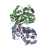
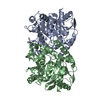
| Structure viewer | Molecule: MolmilJmol/JSmol |
|---|
- Downloads & links
-Download
| PDBx/mmCIF format | 2pnj.cif.gz | 163.5 KB | Display | PDBx/mmCIF format |
|---|---|---|---|---|
| PDB format | pdb2pnj.ent.gz | 129 KB | Display | PDB format |
| PDBx/mmJSON format | 2pnj.json.gz | Tree view | PDBx/mmJSON format | |
| Others |  Other downloads Other downloads |
-Validation report
| Arichive directory | https://data.pdbj.org/pub/pdb/validation_reports/pn/2pnjftp://data.pdbj.org/pub/pdb/validation_reports/pn/2pnj | HTTPS FTP |
|---|
-Related structure data
| Related structure data |  2po5C  2po7C  1hrkS S: Starting model for refinement C: citing same article ( |
|---|---|
| Similar structure data |


-Links
 PDBj
PDBj


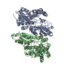
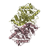
- Assembly
| Deposited unit | 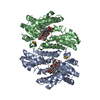
| ||||||||||||||||||
|---|---|---|---|---|---|---|---|---|---|---|---|---|---|---|---|---|---|---|---|
| 1 |
| ||||||||||||||||||
| Unit cell |
| ||||||||||||||||||
| Noncrystallographic symmetry (NCS) | NCS domain:
NCS domain segments: Component-ID: 1 / Ens-ID: 1 / Beg auth comp-ID: ARG / Beg label comp-ID: ARG / End auth comp-ID: LEU / End label comp-ID: LEU / Refine code: 4 / Auth seq-ID: 65 - 423 / Label seq-ID: 1 - 359
|
-Components
| #1: Protein | / E.C.4.99.1.1 / Protoheme ferro-lyase / Heme synthetase Mass: 41058.254 Da / Num. of mol.: 2 / Fragment: Mature Protein / Mutation: R115L, F337A Source method: isolated from a genetically manipulated source Source: (gene. exp.) Homo sapiens (human) / Gene: FECH / Plasmid: PHDTF20 / Production host:  Escherichia coli (E. coli) / Strain (production host): JM109 / References: UniProt: P22830, protoporphyrin ferrochelatase Escherichia coli (E. coli) / Strain (production host): JM109 / References: UniProt: P22830, protoporphyrin ferrochelatase#2: Chemical | Iron–sulfur cluster  Mass: 175.820 Da / Num. of mol.: 2 / Source method: obtained synthetically / Formula: Fe2S2 Mass: 175.820 Da / Num. of mol.: 2 / Source method: obtained synthetically / Formula: Fe2S2#3: Chemical | ChemComp-CHD / Cholic acid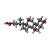  Mass: 408.571 Da / Num. of mol.: 6 / Source method: obtained synthetically / Formula: C24H40O5 Mass: 408.571 Da / Num. of mol.: 6 / Source method: obtained synthetically / Formula: C24H40O5#4: Water | ChemComp-HOH / | Water Mass: 18.015 Da / Num. of mol.: 342 / Source method: isolated from a natural source / Formula: H2O Mass: 18.015 Da / Num. of mol.: 342 / Source method: isolated from a natural source / Formula: H2O |
|---|
-Experimental details
-Experiment
| Experiment | Method: X-RAY DIFFRACTION / Number of used crystals: 1 |
|---|
- Sample preparation
| Crystal | Density Matthews: 2.81 Å3/Da / Density % sol: 56.23 % |
|---|---|
| Crystal grow | Temperature: 291 K / Method: microbatch / pH: 6.5 Details: MICROBATCH UNDER OIL (70:30 PARAFFIN/SILICONE OIL MIXTURE) METHO USING 2 microliter DROPS CONTAINING EQUAL VOLUMES OF PROTEIN SOLUTION (64 MG/ML IN 50 MM TRIS MOPS, 0.1M KCL, 1% NA-CHOLATE, ...Details: MICROBATCH UNDER OIL (70:30 PARAFFIN/SILICONE OIL MIXTURE) METHO USING 2 microliter DROPS CONTAINING EQUAL VOLUMES OF PROTEIN SOLUTION (64 MG/ML IN 50 MM TRIS MOPS, 0.1M KCL, 1% NA-CHOLATE, 250 MM IMIDAZOLE, PH 8.1) AND PRECIPITANT SOLUTION (0.2 M SODIUM ACETAT TRI HYDRATE, 0.1 M SODIUM CACODYLATE, PH 6.5 IN 30%W/V POLYETHYL GLYCOL 8000 (HAMPTON CRYSTAL SCREEN I-28, HAMPTON RESEARCH), pH 6.50, temperature 291K |
-Data collection
| Diffraction | Mean temperature: 100 K | |||||||||
|---|---|---|---|---|---|---|---|---|---|---|
| Diffraction source | Source: SYNCHROTRON / Site: APS  / Beamline: 22-BM / Wavelength: 1 / Wavelength: 1 Å / Beamline: 22-BM / Wavelength: 1 / Wavelength: 1 Å | |||||||||
| Detector | Type: MARMOSAIC 225 mm CCD / Detector: CCD / Date: Nov 18, 2005 / Details: ROSENBAUM | |||||||||
| Radiation | Monochromator: SI 111 / Protocol: SINGLE WAVELENGTH / Monochromatic (M) / Laue (L): M / Scattering type: x-ray | |||||||||
| Radiation wavelength |
| |||||||||
| Reflection | Resolution: 2→50 Å / Num. obs: 58933 / % possible obs: 90.1 % / Redundancy: 17.5 % / Rmerge(I) obs: 0.057 / Net I/σ(I): 25.23 | |||||||||
| Reflection shell | Resolution: 2→2.07 Å / Redundancy: 10 % / Rmerge(I) obs: 0.242 / Mean I/σ(I) obs: 3.39 / % possible all: 98.2 |
- Processing
| Software |
| ||||||||||||||||||||||||||||||||||||||||||||||||||||||||||||||||||||||||||||||||||||||||||
|---|---|---|---|---|---|---|---|---|---|---|---|---|---|---|---|---|---|---|---|---|---|---|---|---|---|---|---|---|---|---|---|---|---|---|---|---|---|---|---|---|---|---|---|---|---|---|---|---|---|---|---|---|---|---|---|---|---|---|---|---|---|---|---|---|---|---|---|---|---|---|---|---|---|---|---|---|---|---|---|---|---|---|---|---|---|---|---|---|---|---|---|
| Refinement | Method to determine structure: DIFFERENCE FOURIER Starting model: 1HRK Resolution: 2.35→43.44 Å / Cor.coef. Fo:Fc: 0.951 / Cor.coef. Fo:Fc free: 0.91 / SU B: 6.661 / SU ML: 0.165 / Cross valid method: THROUGHOUT / σ(F): 0 / σ(I): 0 / ESU R: 0.332 / ESU R Free: 0.251 / Stereochemistry target values: MAXIMUM LIKELIHOOD / Details: HYDROGENS HAVE BEEN ADDED IN THE RIDING POSITIONS
| ||||||||||||||||||||||||||||||||||||||||||||||||||||||||||||||||||||||||||||||||||||||||||
| Solvent computation | Ion probe radii: 0.8 Å / Shrinkage radii: 0.8 Å / VDW probe radii: 1.2 Å / Solvent model: MASK | ||||||||||||||||||||||||||||||||||||||||||||||||||||||||||||||||||||||||||||||||||||||||||
| Displacement parameters | Biso mean: 25.034 Å2
| ||||||||||||||||||||||||||||||||||||||||||||||||||||||||||||||||||||||||||||||||||||||||||
| Refinement step | Cycle: LAST / Resolution: 2.35→43.44 Å
| ||||||||||||||||||||||||||||||||||||||||||||||||||||||||||||||||||||||||||||||||||||||||||
| Refine LS restraints |
| ||||||||||||||||||||||||||||||||||||||||||||||||||||||||||||||||||||||||||||||||||||||||||
| Refine LS restraints NCS | Dom-ID: 1 / Auth asym-ID: A / Ens-ID: 1 / Number: 2811 / Refine-ID: X-RAY DIFFRACTION
| ||||||||||||||||||||||||||||||||||||||||||||||||||||||||||||||||||||||||||||||||||||||||||
| LS refinement shell | Resolution: 2.35→2.411 Å / Total num. of bins used: 20
|
