 Movie
Movie Controller

[English] 日本語
 Yorodumi
- PDB-2hre: Structure of human ferrochelatase variant E343K with protoporphyr... -
+ Open data

- Basic information
| Entry | Database: PDB / ID: 2hre | ||||||
|---|---|---|---|---|---|---|---|
| Title | Structure of human ferrochelatase variant E343K with protoporphyrin IX bound | ||||||
 Components Components | Ferrochelatase | ||||||
 Keywords Keywords | LYASE / Heme synthesis / Ferrochelatase / Protoporphyrin IX | ||||||
| Function / homology |  Function and homology information Function and homology informationprotoporphyrinogen IX metabolic process / regulation of hemoglobin biosynthetic process / regulation of eIF2 alpha phosphorylation by heme / detection of UV / iron-responsive element binding / response to platinum ion / protoporphyrin ferrochelatase / response to insecticide / heme B biosynthetic process / heme O biosynthetic process ...protoporphyrinogen IX metabolic process / regulation of hemoglobin biosynthetic process / regulation of eIF2 alpha phosphorylation by heme / detection of UV / iron-responsive element binding / response to platinum ion / protoporphyrin ferrochelatase / response to insecticide / heme B biosynthetic process / heme O biosynthetic process / ferrochelatase activity / heme A biosynthetic process / very-low-density lipoprotein particle assembly / response to methylmercury / response to arsenic-containing substance / Heme biosynthesis / heme biosynthetic process / response to light stimulus / cholesterol metabolic process / cellular response to dexamethasone stimulus / erythrocyte differentiation / generation of precursor metabolites and energy / ferrous iron binding / response to lead ion / 2 iron, 2 sulfur cluster binding / multicellular organismal-level iron ion homeostasis / response to ethanol / mitochondrial inner membrane / mitochondrial matrix / response to xenobiotic stimulus / heme binding / protein homodimerization activity / mitochondrion / identical protein bindingSimilarity search - Function | ||||||
| Biological species |  Homo sapiens (human) Homo sapiens (human) | ||||||
| Method | X-RAY DIFFRACTION / MOLECULAR REPLACEMENT / Resolution: 2.5 Å | ||||||
 Authors Authors | Medlock, A. / Swartz, L. / Dailey, T.A. / Dailey, H.A. / Lanzilotta, W.N. | ||||||
 Citation Citation | Journal: Proc.Natl.Acad.Sci.Usa / Year: 2007 Title: Substrate interactions with human ferrochelatase Authors: Medlock, A. / Swartz, L. / Dailey, T.A. / Dailey, H.A. / Lanzilotta, W.N. | ||||||
| History |
|
- Structure visualization
| Structure viewer | Molecule: MolmilJmol/JSmol |
|---|
- Downloads & links
-Download
| PDBx/mmCIF format | 2hre.cif.gz | 307.2 KB | Display | PDBx/mmCIF format |
|---|---|---|---|---|
| PDB format | pdb2hre.ent.gz | 250.4 KB | Display | PDB format |
| PDBx/mmJSON format | 2hre.json.gz | Tree view | PDBx/mmJSON format | |
| Others |  Other downloads Other downloads |
-Validation report
| Arichive directory | https://data.pdbj.org/pub/pdb/validation_reports/hr/2hreftp://data.pdbj.org/pub/pdb/validation_reports/hr/2hre | HTTPS FTP |
|---|
-Related structure data
| Related structure data |  2hrcC  1hrkS C: citing same article ( S: Starting model for refinement |
|---|---|
| Similar structure data |


-Links
 PDBj
PDBj


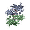
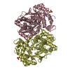
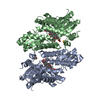
- Assembly
| Deposited unit | 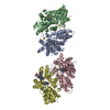
| ||||||||
|---|---|---|---|---|---|---|---|---|---|
| 1 | 
| ||||||||
| 2 | 
| ||||||||
| 3 | 
| ||||||||
| Unit cell |
| ||||||||
| Details | Their are two biologial units in the asymmetric unit. The first biological unit (dimer) is formed by monomers A and B. The second biological unti is formed by monomer C and the symmetry mate of D. |
-Components
| #1: Protein | / Protoheme ferro-lyase / Heme synthetase Mass: 41178.453 Da / Num. of mol.: 4 / Mutation: E343K Source method: isolated from a genetically manipulated source Source: (gene. exp.) Homo sapiens (human) / Description: PLASMID / Gene: FECH / Plasmid: pTRCHis / Production host:  Escherichia coli (E. coli) / References: UniProt: P22830, protoporphyrin ferrochelatase Escherichia coli (E. coli) / References: UniProt: P22830, protoporphyrin ferrochelatase#2: Chemical | ChemComp-PP9 / Protoporphyrin IX  Mass: 562.658 Da / Num. of mol.: 6 / Source method: obtained synthetically / Formula: C34H34N4O4 Mass: 562.658 Da / Num. of mol.: 6 / Source method: obtained synthetically / Formula: C34H34N4O4#3: Chemical | ChemComp-FES / Iron–sulfur cluster  Mass: 175.820 Da / Num. of mol.: 4 / Source method: obtained synthetically / Formula: Fe2S2 Mass: 175.820 Da / Num. of mol.: 4 / Source method: obtained synthetically / Formula: Fe2S2#4: Chemical | Cholic acid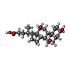  Mass: 408.571 Da / Num. of mol.: 2 / Source method: obtained synthetically / Formula: C24H40O5 Mass: 408.571 Da / Num. of mol.: 2 / Source method: obtained synthetically / Formula: C24H40O5#5: Water | ChemComp-HOH / | Water Mass: 18.015 Da / Num. of mol.: 338 / Source method: isolated from a natural source / Formula: H2O Mass: 18.015 Da / Num. of mol.: 338 / Source method: isolated from a natural source / Formula: H2O |
|---|
-Experimental details
-Experiment
| Experiment | Method: X-RAY DIFFRACTION / Number of used crystals: 1 |
|---|
- Sample preparation
| Crystal | Density Matthews: 2.65 Å3/Da / Density % sol: 53.66 % |
|---|---|
| Crystal grow | Temperature: 298 K / Method: vapor diffusion, hanging drop / pH: 5.5 Details: 0.1 M Bis-Tris, 0.2 M Magnesium Chloride, 25% PEG 3350, pH 5.5, VAPOR DIFFUSION, HANGING DROP, temperature 298K |
-Data collection
| Diffraction | Mean temperature: 120 K |
|---|---|
| Diffraction source | Source: ROTATING ANODE / Type: RIGAKU / Wavelength: 1.5418 Å |
| Detector | Type: SIEMENS / Detector: CCD / Date: Dec 15, 2005 / Details: Osmic Blue |
| Radiation | Monochromator: Copper FRD / Protocol: SINGLE WAVELENGTH / Monochromatic (M) / Laue (L): M / Scattering type: x-ray |
| Radiation wavelength | Wavelength: 1.5418 Å / Relative weight: 1 |
| Reflection | Resolution: 2.5→40 Å / Num. all: 58659 / Num. obs: 53845 / % possible obs: 96.7 % / Observed criterion σ(F): 2 / Observed criterion σ(I): 2 / Biso Wilson estimate: 23.2 Å2 / Rmerge(I) obs: 0.072 / Rsym value: 0.08 / Net I/σ(I): 13.5 |
| Reflection shell | Resolution: 2.5→2.59 Å / Redundancy: 4 % / Rmerge(I) obs: 0.281 / Mean I/σ(I) obs: 3.4 / Num. unique all: 53845 / Rsym value: 0.322 / % possible all: 93.5 |
- Processing
| Software |
| |||||||||||||||||||||||||
|---|---|---|---|---|---|---|---|---|---|---|---|---|---|---|---|---|---|---|---|---|---|---|---|---|---|---|
| Refinement | Method to determine structure: MOLECULAR REPLACEMENT Starting model: PDB ENTRY 1HRK Resolution: 2.5→43.66 Å / Rfactor Rfree error: 0.005 / Data cutoff high absF: 400690.97 / Data cutoff low absF: 0 / Isotropic thermal model: RESTRAINED / Cross valid method: THROUGHOUT / σ(F): 0 / Stereochemistry target values: Engh & Huber
| |||||||||||||||||||||||||
| Solvent computation | Solvent model: FLAT MODEL / Bsol: 48.4191 Å2 / ksol: 0.398897 e/Å3 | |||||||||||||||||||||||||
| Displacement parameters | Biso mean: 31.1 Å2
| |||||||||||||||||||||||||
| Refine analyze |
| |||||||||||||||||||||||||
| Refinement step | Cycle: LAST / Resolution: 2.5→43.66 Å
| |||||||||||||||||||||||||
| Refine LS restraints |
| |||||||||||||||||||||||||
| LS refinement shell | Resolution: 2.5→2.66 Å / Rfactor Rfree error: 0.018 / Total num. of bins used: 6
| |||||||||||||||||||||||||
| Xplor file |
|
