 Movie
Movie Controller

[English] 日本語
 Yorodumi
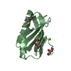
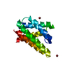
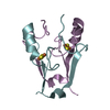
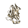
- PDB-6nv1: Structure of drug-resistant V27A mutant of the influenza M2 proto... -
+ Open data

- Basic information
| Entry | Database: PDB / ID: 6nv1 | ||||||
|---|---|---|---|---|---|---|---|
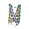
| Title | Structure of drug-resistant V27A mutant of the influenza M2 proton channel bound to spiroadamantyl amine inhibitor | ||||||
 Components Components | Matrix protein 2 | ||||||
 Keywords Keywords |  MEMBRANE PROTEIN / proton channel MEMBRANE PROTEIN / proton channel | ||||||
| Function / homology |  Function and homology information Function and homology informationsuppression by virus of host autophagy / : / proton transmembrane transporter activity / : / protein complex oligomerization / monoatomic ion channel activity / membrane => GO:0016020 / host cell plasma membrane / virion membrane Similarity search - Function | ||||||
| Biological species |   Influenza A virus Influenza A virus | ||||||
| Method | X-RAY DIFFRACTION / SYNCHROTRON / MOLECULAR REPLACEMENT / Resolution: 2.5 Å | ||||||
 Authors Authors | Thomaston, J.L. / Liu, L. / DeGrado, W.F. | ||||||
| Funding support |  United States, 1items United States, 1items
| ||||||
 Citation Citation | Journal: Biochemistry / Year: 2020 Title: X-ray Crystal Structures of the Influenza M2 Proton Channel Drug-Resistant V27A Mutant Bound to a Spiro-Adamantyl Amine Inhibitor Reveal the Mechanism of Adamantane Resistance. Authors: Thomaston, J.L. / Konstantinidi, A. / Liu, L. / Lambrinidis, G. / Tan, J. / Caffrey, M. / Wang, J. / Degrado, W.F. / Kolocouris, A. | ||||||
| History |
|
- Structure visualization
| Structure viewer | Molecule: MolmilJmol/JSmol |
|---|
- Downloads & links
-Download
| PDBx/mmCIF format | 6nv1.cif.gz | 54.6 KB | Display | PDBx/mmCIF format |
|---|---|---|---|---|
| PDB format | pdb6nv1.ent.gz | 40.3 KB | Display | PDB format |
| PDBx/mmJSON format | 6nv1.json.gz | Tree view | PDBx/mmJSON format | |
| Others |  Other downloads Other downloads |
-Validation report
| Arichive directory | https://data.pdbj.org/pub/pdb/validation_reports/nv/6nv1ftp://data.pdbj.org/pub/pdb/validation_reports/nv/6nv1 | HTTPS FTP |
|---|
-Related structure data
| Related structure data | 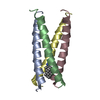 6ougC 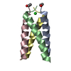 6bkkS S: Starting model for refinement C: citing same article ( |
|---|---|
| Similar structure data |


-Links
 PDBj
PDBj


- Assembly
| Deposited unit | 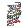
| ||||||||
|---|---|---|---|---|---|---|---|---|---|
| 1 | 
| ||||||||
| 2 | 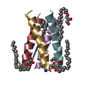
| ||||||||
| Unit cell |
|
-Components
| #1: Protein/peptide | Mass: 2726.287 Da / Num. of mol.: 8 / Source method: obtained synthetically / Source: (synth.) Influenza A virus / References: UniProt: A4D7H3#2: Chemical | ChemComp-OLC / (   Mass: 356.540 Da / Num. of mol.: 8 / Source method: obtained synthetically / Formula: C21H40O4 Mass: 356.540 Da / Num. of mol.: 8 / Source method: obtained synthetically / Formula: C21H40O4#3: Chemical | 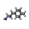  Mass: 219.366 Da / Num. of mol.: 2 / Source method: obtained synthetically / Formula: C15H25N Mass: 219.366 Da / Num. of mol.: 2 / Source method: obtained synthetically / Formula: C15H25N#4: Chemical | Chloride  Mass: 35.453 Da / Num. of mol.: 2 / Source method: obtained synthetically / Formula: Cl Mass: 35.453 Da / Num. of mol.: 2 / Source method: obtained synthetically / Formula: Cl#5: Water | ChemComp-HOH / | Water Mass: 18.015 Da / Num. of mol.: 30 / Source method: isolated from a natural source / Formula: H2O Mass: 18.015 Da / Num. of mol.: 30 / Source method: isolated from a natural source / Formula: H2O |
|---|
-Experimental details
-Experiment
| Experiment | Method: X-RAY DIFFRACTION / Number of used crystals: 1 |
|---|
- Sample preparation
| Crystal | Density Matthews: 2.31 Å3/Da / Density % sol: 46.73 % |
|---|---|
| Crystal grow | Temperature: 300 K / Method: lipidic cubic phase Details: 0.045 M HEPES pH 7.5, 19.8% w/v PEG 4000, 0.01 M L-proline, monoolein, MNG-34, spiroadamantyl amine |
-Data collection
| Diffraction | Mean temperature: 100 K / Serial crystal experiment: N |
|---|---|
| Diffraction source | Source: SYNCHROTRON / Site: ALS / Beamline: 8.3.1 / Wavelength: 1.1158 Å |
| Detector | Type: DECTRIS PILATUS3 S 6M / Detector: PIXEL / Date: Dec 20, 2016 |
| Radiation | Protocol: SINGLE WAVELENGTH / Monochromatic (M) / Laue (L): M / Scattering type: x-ray |
| Radiation wavelength | Wavelength: 1.1158 Å / Relative weight: 1 |
| Reflection | Resolution: 2.5→41.58 Å / Num. obs: 6840 / % possible obs: 99.25 % / Redundancy: 10.3 % / CC1/2: 1 / Rmerge(I) obs: 0.1262 / Net I/σ(I): 13.27 |
| Reflection shell | Resolution: 2.5→2.589 Å / Redundancy: 3.52 % / Rmerge(I) obs: 0.7294 / Mean I/σ(I) obs: 3.52 / Num. unique obs: 653 / CC1/2: 0.941 / % possible all: 98.93 |
- Processing
| Software |
| ||||||||||||||||||||||||||||||||||||||||||
|---|---|---|---|---|---|---|---|---|---|---|---|---|---|---|---|---|---|---|---|---|---|---|---|---|---|---|---|---|---|---|---|---|---|---|---|---|---|---|---|---|---|---|---|
| Refinement | Method to determine structure: MOLECULAR REPLACEMENT Starting model: 6BKK Resolution: 2.5→41.579 Å / SU ML: 0.28 / Cross valid method: FREE R-VALUE / σ(F): 1.34 / Phase error: 33.38 / Stereochemistry target values: ML
| ||||||||||||||||||||||||||||||||||||||||||
| Solvent computation | Shrinkage radii: 0.9 Å / VDW probe radii: 1.11 Å / Solvent model: FLAT BULK SOLVENT MODEL | ||||||||||||||||||||||||||||||||||||||||||
| Refinement step | Cycle: LAST / Resolution: 2.5→41.579 Å
| ||||||||||||||||||||||||||||||||||||||||||
| Refine LS restraints |
| ||||||||||||||||||||||||||||||||||||||||||
| LS refinement shell |
|
