 Movie
Movie Controller


+ Open data

- Basic information
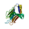
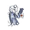
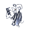
| Entry | Database: PDB / ID: 2yps | ||||||
|---|---|---|---|---|---|---|---|
| Title | Crystal structure of the PX domain of human sorting nexin 3 | ||||||
 Components Components | SORTING NEXIN-3 | ||||||
 Keywords Keywords | PROTEIN TRANSPORT / ENDOSOME | ||||||
| Function / homology |  Function and homology information Function and homology informationnegative regulation of early endosome to late endosome transport / late endosome to Golgi transport / protein to membrane docking / negative regulation of protein transport / membrane invagination / WNT ligand biogenesis and trafficking / intralumenal vesicle formation / retromer complex binding / phosphatidylinositol-5-phosphate binding / retromer complex ...negative regulation of early endosome to late endosome transport / late endosome to Golgi transport / protein to membrane docking / negative regulation of protein transport / membrane invagination / WNT ligand biogenesis and trafficking / intralumenal vesicle formation / retromer complex binding / phosphatidylinositol-5-phosphate binding / retromer complex / negative regulation of viral entry into host cell / phosphatidylinositol-3-phosphate binding / early phagosome / endocytic recycling / regulation of Wnt signaling pathway / phosphatidylinositol-4-phosphate binding / phosphatidylinositol-3,5-bisphosphate binding / negative regulation of phagocytosis / clathrin-coated vesicle / response to bacterium / negative regulation of protein catabolic process / positive regulation of neuron projection development / protein transport / early endosome membrane / protein phosphatase binding / early endosome / endosome membrane / Ub-specific processing proteases / extracellular exosome / cytosol / cytoplasmSimilarity search - Function | ||||||
| Biological species |  HOMO SAPIENS (human) HOMO SAPIENS (human) | ||||||
| Method | X-RAY DIFFRACTION / SYNCHROTRON / MOLECULAR REPLACEMENT / Resolution: 2.6 Å | ||||||
 Authors Authors | Canning, P. / Kiyani, W. / Froese, D.S. / Krojer, T. / Strain-Damerell, C. / von Delft, F. / Arrowsmith, C.H. / Edwards, A.M. / Bountra, C. / Yue, W.W. | ||||||
 Citation Citation | Journal: To be Published Title: Crystal Structure of the Px Domain of Human Sorting Nexin 3 Authors: Canning, P. / Froese, D.S. / Krojer, T. / Strain-Damerell, C. / von Delft, F. / Arrowsmith, C.H. / Edwards, A.M. / Bountra, C. / Yue, W.W. | ||||||
| History |
|
- Structure visualization
| Structure viewer | Molecule: MolmilJmol/JSmol |
|---|
- Downloads & links
-Download
| PDBx/mmCIF format | 2yps.cif.gz | 168.7 KB | Display | PDBx/mmCIF format |
|---|---|---|---|---|
| PDB format | pdb2yps.ent.gz | 132.9 KB | Display | PDB format |
| PDBx/mmJSON format | 2yps.json.gz | Tree view | PDBx/mmJSON format | |
| Others |  Other downloads Other downloads |
-Validation report
| Arichive directory | https://data.pdbj.org/pub/pdb/validation_reports/yp/2ypsftp://data.pdbj.org/pub/pdb/validation_reports/yp/2yps | HTTPS FTP |
|---|
-Related structure data
| Related structure data | 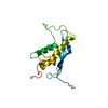 2cskS S: Starting model for refinement |
|---|---|
| Similar structure data |


-Links
 PDBj
PDBj- Assembly
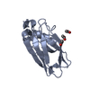
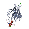
| Deposited unit | 
| ||||||||||||||||||||||||||||
|---|---|---|---|---|---|---|---|---|---|---|---|---|---|---|---|---|---|---|---|---|---|---|---|---|---|---|---|---|---|
| 1 | 
| ||||||||||||||||||||||||||||
| 2 | 
| ||||||||||||||||||||||||||||
| 3 | 
| ||||||||||||||||||||||||||||
| 4 | 
| ||||||||||||||||||||||||||||
| Unit cell |
| ||||||||||||||||||||||||||||
| Components on special symmetry positions |
| ||||||||||||||||||||||||||||
| Noncrystallographic symmetry (NCS) | NCS oper:
|
-Components
| #1: Protein | / PROTEIN SDP3 Mass: 15652.799 Da / Num. of mol.: 4 / Fragment: PX DOMAIN, RESIDUES 24-155 Source method: isolated from a genetically manipulated source Source: (gene. exp.) HOMO SAPIENS (human) / Description: MGC / Plasmid: PNIC28-BSA4 / Production host:  ESCHERICHIA COLI (E. coli) / Strain (production host): BL21(DE3) / Variant (production host): R3-PRARE2 / References: UniProt: O60493 ESCHERICHIA COLI (E. coli) / Strain (production host): BL21(DE3) / Variant (production host): R3-PRARE2 / References: UniProt: O60493#2: Water | ChemComp-HOH / | Water Mass: 18.015 Da / Num. of mol.: 10 / Source method: isolated from a natural source / Formula: H2O Mass: 18.015 Da / Num. of mol.: 10 / Source method: isolated from a natural source / Formula: H2O |
|---|
-Experimental details
-Experiment
| Experiment | Method: X-RAY DIFFRACTION / Number of used crystals: 1 |
|---|
- Sample preparation
| Crystal | Density Matthews: 3.54 Å3/Da / Density % sol: 65.3 % / Description: NONE |
|---|---|
| Crystal grow | Details: 3.5M FORMATE |
-Data collection
| Diffraction | Mean temperature: 100 K |
|---|---|
| Diffraction source | Source: SYNCHROTRON / Site: Diamond  / Beamline: I04 / Wavelength: 0.9611 / Beamline: I04 / Wavelength: 0.9611 |
| Detector | Type: ADSC CCD / Detector: CCD / Date: Sep 29, 2012 |
| Radiation | Protocol: SINGLE WAVELENGTH / Monochromatic (M) / Laue (L): M / Scattering type: x-ray |
| Radiation wavelength | Wavelength: 0.9611 Å / Relative weight: 1 |
| Reflection | Resolution: 2.6→51.02 Å / Num. obs: 16601 / % possible obs: 99.7 % / Observed criterion σ(I): 2 / Redundancy: 3.5 % / Rmerge(I) obs: 0.08 / Net I/σ(I): 8.2 |
| Reflection shell | Resolution: 2.6→2.73 Å / Redundancy: 3.6 % / Rmerge(I) obs: 0.5 / Mean I/σ(I) obs: 2.1 / % possible all: 100 |
- Processing
| Software |
| ||||||||||||||||||||||||||||||||||||||||||||||||||||||||||||||||||||||||||||||||||||||||||||||||||||||||||||||||||||||||||||||||||||||||||||||||||||||||||||||||||||||||||||||||||||||
|---|---|---|---|---|---|---|---|---|---|---|---|---|---|---|---|---|---|---|---|---|---|---|---|---|---|---|---|---|---|---|---|---|---|---|---|---|---|---|---|---|---|---|---|---|---|---|---|---|---|---|---|---|---|---|---|---|---|---|---|---|---|---|---|---|---|---|---|---|---|---|---|---|---|---|---|---|---|---|---|---|---|---|---|---|---|---|---|---|---|---|---|---|---|---|---|---|---|---|---|---|---|---|---|---|---|---|---|---|---|---|---|---|---|---|---|---|---|---|---|---|---|---|---|---|---|---|---|---|---|---|---|---|---|---|---|---|---|---|---|---|---|---|---|---|---|---|---|---|---|---|---|---|---|---|---|---|---|---|---|---|---|---|---|---|---|---|---|---|---|---|---|---|---|---|---|---|---|---|---|---|---|---|---|
| Refinement | Method to determine structure: MOLECULAR REPLACEMENT Starting model: PDB ENTRY 2CSK Resolution: 2.6→51.02 Å / Cor.coef. Fo:Fc: 0.934 / Cor.coef. Fo:Fc free: 0.894 / SU B: 20.64 / SU ML: 0.205 / Cross valid method: THROUGHOUT / ESU R: 0.54 / ESU R Free: 0.3 / Stereochemistry target values: MAXIMUM LIKELIHOOD / Details: HYDROGENS HAVE BEEN ADDED IN THE RIDING POSITIONS.
| ||||||||||||||||||||||||||||||||||||||||||||||||||||||||||||||||||||||||||||||||||||||||||||||||||||||||||||||||||||||||||||||||||||||||||||||||||||||||||||||||||||||||||||||||||||||
| Solvent computation | Ion probe radii: 0.8 Å / Shrinkage radii: 0.8 Å / VDW probe radii: 1.2 Å / Solvent model: MASK | ||||||||||||||||||||||||||||||||||||||||||||||||||||||||||||||||||||||||||||||||||||||||||||||||||||||||||||||||||||||||||||||||||||||||||||||||||||||||||||||||||||||||||||||||||||||
| Displacement parameters | Biso mean: 48.029 Å2
| ||||||||||||||||||||||||||||||||||||||||||||||||||||||||||||||||||||||||||||||||||||||||||||||||||||||||||||||||||||||||||||||||||||||||||||||||||||||||||||||||||||||||||||||||||||||
| Refinement step | Cycle: LAST / Resolution: 2.6→51.02 Å
| ||||||||||||||||||||||||||||||||||||||||||||||||||||||||||||||||||||||||||||||||||||||||||||||||||||||||||||||||||||||||||||||||||||||||||||||||||||||||||||||||||||||||||||||||||||||
| Refine LS restraints |
|