 Movie
Movie Controller

[English] 日本語
 Yorodumi
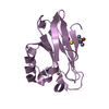
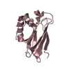
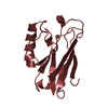
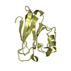
- PDB-4hz1: Crystal Structure of Pseudomonas aeruginosa azurin with iron(II) ... -
+ Open data

- Basic information
| Entry | Database: PDB / ID: 4hz1 | ||||||
|---|---|---|---|---|---|---|---|
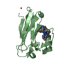
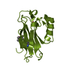
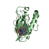
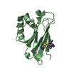
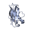
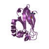
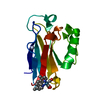
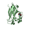
| Title | Crystal Structure of Pseudomonas aeruginosa azurin with iron(II) at the copper-binding site. | ||||||
 Components Components | Azurin | ||||||
 Keywords Keywords | ELECTRON TRANSPORT / Copper binding site / reductase / arsenite | ||||||
| Function / homology |  Function and homology information Function and homology informationtransition metal ion binding / electron transfer activity / periplasmic space / copper ion binding / zinc ion binding / identical protein bindingSimilarity search - Function | ||||||
| Biological species |   Pseudomonas aeruginosa (bacteria) Pseudomonas aeruginosa (bacteria) | ||||||
| Method | X-RAY DIFFRACTION / SYNCHROTRON / MOLECULAR REPLACEMENT / Resolution: 2.2 Å | ||||||
 Authors Authors | McLaughlin, M.P. / Retegan, M. / Bill, E. / Payne, T.M. / Shafaat, H.S. / Pea, S. / Sudhamsu, J. / Ensign, A.A. / Crane, B.R. / Neese, F. / Holland, P.L. | ||||||
 Citation Citation | Journal: J.Am.Chem.Soc. / Year: 2012 Title: Azurin as a Protein Scaffold for a Low-coordinate Nonheme Iron Site with a Small-molecule Binding Pocket. Authors: McLaughlin, M.P. / Retegan, M. / Bill, E. / Payne, T.M. / Shafaat, H.S. / Pena, S. / Sudhamsu, J. / Ensign, A.A. / Crane, B.R. / Neese, F. / Holland, P.L. | ||||||
| History |
|
- Structure visualization
| Structure viewer | Molecule: MolmilJmol/JSmol |
|---|
- Downloads & links
-Download
| PDBx/mmCIF format | 4hz1.cif.gz | 109.8 KB | Display | PDBx/mmCIF format |
|---|---|---|---|---|
| PDB format | pdb4hz1.ent.gz | 84.5 KB | Display | PDB format |
| PDBx/mmJSON format | 4hz1.json.gz | Tree view | PDBx/mmJSON format | |
| Others |  Other downloads Other downloads |
-Validation report
| Arichive directory | https://data.pdbj.org/pub/pdb/validation_reports/hz/4hz1ftp://data.pdbj.org/pub/pdb/validation_reports/hz/4hz1 | HTTPS FTP |
|---|
-Related structure data
| Related structure data |  1azuS S: Starting model for refinement |
|---|---|
| Similar structure data |


-Links
 PDBj
PDBj


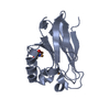
- Assembly
| Deposited unit | 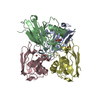
| ||||||||
|---|---|---|---|---|---|---|---|---|---|
| 1 | 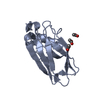
| ||||||||
| 2 | 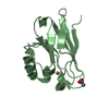
| ||||||||
| 3 | 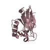
| ||||||||
| 4 | 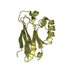
| ||||||||
| Unit cell |
|
-Components
| #1: Protein | Mass: 13961.799 Da / Num. of mol.: 4 Source method: isolated from a genetically manipulated source Source: (gene. exp.) Pseudomonas aeruginosa (bacteria) / Strain: ATCC 15692 / PAO1 / 1C / PRS 101 / LMG 12228 / Gene: azu, PA4922 / Plasmid: pET9a / Production host: Escherichia coli (E. coli) / Strain (production host): BL21(DE3) / References: UniProt: P00282#2: Chemical | ChemComp-ACT / Acetate  Mass: 59.044 Da / Num. of mol.: 5 / Source method: obtained synthetically / Formula: C2H3O2 Mass: 59.044 Da / Num. of mol.: 5 / Source method: obtained synthetically / Formula: C2H3O2#3: Chemical |   Mass: 55.845 Da / Num. of mol.: 2 / Source method: obtained synthetically / Formula: Fe Mass: 55.845 Da / Num. of mol.: 2 / Source method: obtained synthetically / Formula: Fe#4: Water | ChemComp-HOH / | Water Mass: 18.015 Da / Num. of mol.: 147 / Source method: isolated from a natural source / Formula: H2O Mass: 18.015 Da / Num. of mol.: 147 / Source method: isolated from a natural source / Formula: H2O |
|---|
-Experimental details
-Experiment
| Experiment | Method: X-RAY DIFFRACTION / Number of used crystals: 1 |
|---|
- Sample preparation
| Crystal | Density Matthews: 2.26 Å3/Da / Density % sol: 45.6 % |
|---|---|
| Crystal grow | Temperature: 300 K / Method: vapor diffusion, hanging drop Details: 3.0 - 3.5 M ammonium sulfate, 0.5 M lithium nitrate, 0.1 M sodium acetate pH 5.2-5.4, VAPOR DIFFUSION, HANGING DROP, temperature 300K PH range: 5.2-5.4 |
-Data collection
| Diffraction | Mean temperature: 77 K |
|---|---|
| Diffraction source | Source: SYNCHROTRON / Site: CHESS  / Beamline: A1 / Wavelength: 1.608 Å / Beamline: A1 / Wavelength: 1.608 Å |
| Detector | Type: ADSC QUANTUM 210 / Detector: CCD / Date: Mar 15, 2012 |
| Radiation | Protocol: SINGLE WAVELENGTH / Monochromatic (M) / Laue (L): M / Scattering type: x-ray |
| Radiation wavelength | Wavelength: 1.608 Å / Relative weight: 1 |
| Reflection | Resolution: 2.2→43 Å / Num. all: 26498 / Num. obs: 26498 / % possible obs: 95 % / Observed criterion σ(F): 10000 / Observed criterion σ(I): 10000 / Rsym value: 0.127 / Net I/σ(I): 13.48 |
- Processing
| Software |
| ||||||||||||||||||||
|---|---|---|---|---|---|---|---|---|---|---|---|---|---|---|---|---|---|---|---|---|---|
| Refinement | Method to determine structure: MOLECULAR REPLACEMENT Starting model: PDB ENTRY 1AZU Resolution: 2.2→43 Å / σ(F): 10000 / Stereochemistry target values: Engh & Huber
| ||||||||||||||||||||
| Refinement step | Cycle: LAST / Resolution: 2.2→43 Å
|
