[English] 日本語
Yorodumi
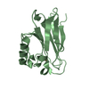
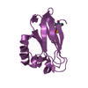
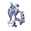
- PDB-4k9j: Structure of Re(CO)3(4,7-dimethyl-phen)(Thr126His)(Lys122Trp)(His... -

+
Open data


ID or keywords:

Loading...

-
Basic information

Entry
Database: PDB / ID: 4k9j
Title
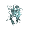
Structure of Re(CO)3(4,7-dimethyl-phen)(Thr126His)(Lys122Trp)(His83Glu)(Trp48Phe)(Tyr72Phe)(Tyr108Phe)AzCu(II), a Rhenium modified Azurin mutant
ComponentsAzurin
KeywordsELECTRON TRANSPORT / rhenium
Function / homology
Function and homology information


transition metal ion binding / periplasmic space / electron transfer activity / copper ion binding / zinc ion binding / identical protein binding
Similarity search - Function
Azurin / Blue (type 1) copper domain / Copper binding proteins, plastocyanin/azurin family / Blue (type 1) copper protein, binding site / Type-1 copper (blue) proteins signature. / Cupredoxins - blue copper proteins / Cupredoxin / Immunoglobulin-like / Sandwich / Mainly Beta
Similarity search - Domain/homology
COPPER (II) ION / Chem-REQ / Azurin
Similarity search - Component
Biological speciesPseudomonas aeruginosa (bacteria)
MethodX-RAY DIFFRACTION / SYNCHROTRON / MOLECULAR REPLACEMENT / Resolution: 1.7 Å
AuthorsTakematsu, K. / Williamson, H.R. / Blanco-Rodriguez, A.M. / Sokolova, L. / Nikolovski, P. / Kaiser, J.T. / Towrie, M. / Clark, I.P. / Vlcek Jr, A. / Winkler, J.R. / Gray, H.B.
CitationJournal: J.Am.Chem.Soc. / Year: 2013
Title: Tryptophan-accelerated electron flow across a protein-protein interface.
Authors: Takematsu, K. / Williamson, H. / Blanco-Rodriguez, A.M. / Sokolova, L. / Nikolovski, P. / Kaiser, J.T. / Towrie, M. / Clark, I.P. / Vlcek, A. / Winkler, J.R. / Gray, H.B.
History
DepositionApr 20, 2013Deposition site: RCSB / Processing site: RCSB
Revision 1.0Oct 2, 2013Provider: repository / Type: Initial release
Revision 1.1Feb 5, 2014Group: Database references
Revision 1.2Sep 20, 2023Group: Data collection / Database references ...Data collection / Database references / Derived calculations / Refinement description
Category: chem_comp_atom / chem_comp_bond ...chem_comp_atom / chem_comp_bond / database_2 / pdbx_initial_refinement_model / pdbx_struct_conn_angle / struct_conn / struct_ref_seq_dif / struct_site
Item: _database_2.pdbx_DOI / _database_2.pdbx_database_accession ..._database_2.pdbx_DOI / _database_2.pdbx_database_accession / _pdbx_struct_conn_angle.ptnr1_auth_comp_id / _pdbx_struct_conn_angle.ptnr1_auth_seq_id / _pdbx_struct_conn_angle.ptnr1_label_atom_id / _pdbx_struct_conn_angle.ptnr1_label_comp_id / _pdbx_struct_conn_angle.ptnr1_label_seq_id / _pdbx_struct_conn_angle.ptnr3_auth_comp_id / _pdbx_struct_conn_angle.ptnr3_auth_seq_id / _pdbx_struct_conn_angle.ptnr3_label_atom_id / _pdbx_struct_conn_angle.ptnr3_label_comp_id / _pdbx_struct_conn_angle.ptnr3_label_seq_id / _pdbx_struct_conn_angle.value / _struct_conn.pdbx_dist_value / _struct_conn.ptnr1_auth_comp_id / _struct_conn.ptnr1_auth_seq_id / _struct_conn.ptnr1_label_atom_id / _struct_conn.ptnr1_label_comp_id / _struct_conn.ptnr1_label_seq_id / _struct_conn.ptnr2_auth_comp_id / _struct_conn.ptnr2_auth_seq_id / _struct_conn.ptnr2_label_asym_id / _struct_conn.ptnr2_label_atom_id / _struct_conn.ptnr2_label_comp_id / _struct_ref_seq_dif.details / _struct_site.pdbx_auth_asym_id / _struct_site.pdbx_auth_comp_id / _struct_site.pdbx_auth_seq_id
Revision 1.3Mar 13, 2024Group: Source and taxonomy / Structure summary / Category: entity / pdbx_entity_src_syn / Item: _entity.details

-
Structure visualization

Structure viewerMolecule:
MolmilJmol/JSmol

Downloads & links

-
Assembly

Deposited unit
A: Azurin
hetero molecules


Theoretical massNumber of molelcules
Total (without water)14,5173
Polymers13,9751
Non-polymers5422
Water1,33374
1


  • Idetical with deposited unit
  • defined by author&software
TypeNameSymmetry operationNumber
identity operation1_555x,y,z1
MethodPISA
Unit cell
Length a, b, c (Å)42.391, 93.215, 109.383
Angle α, β, γ (deg.)90.00, 90.00, 90.00
Int Tables number22
Space group name H-MF222
Components on special symmetry positions
IDModelComponents
11A-364-

HOH

-
Components

#1: Protein Azurin /


Mass: 13974.816 Da / Num. of mol.: 1 / Mutation: t126H, k122w, h83e,w48f, y72f
Source method: isolated from a genetically manipulated source
Source: (gene. exp.) Pseudomonas aeruginosa (bacteria) / Strain: ATCC 15692 / PAO1 / 1C / PRS 101 / LMG 12228 / Gene: azu, PA4922 / Production host: Escherichia coli (E. coli) / Strain (production host): BL21(DE3) / References: UniProt: P00282
#2: Chemical ChemComp-CU / COPPER (II) ION / Copper


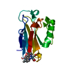
Mass: 63.546 Da / Num. of mol.: 1 / Source method: obtained synthetically / Formula: Cu
#3: Chemical ChemComp-REQ / (1,10 PHENANTHROLINE)-(TRI-CARBON MONOXIDE) RHENIUM (I)


Mass: 478.496 Da / Num. of mol.: 1 / Source method: obtained synthetically / Formula: C17H12N2O3Re
Details: Synthesis described in: Sullivan, B. P.; Meyer, T. J. J. Chem. Soc., Chem. Commun. 1984, 1244-1245. Connick, W. B.; Di Bilio, A. J.; Schaefer, W. P.; Gray, H. B. Acta Crystallogr. C 1999, C55, 913-916
#4: Water ChemComp-HOH / water / Water


Mass: 18.015 Da / Num. of mol.: 74 / Source method: isolated from a natural source / Formula: H2O

-
Experimental details

-
Experiment

ExperimentMethod: X-RAY DIFFRACTION / Number of used crystals: 1

-
Sample preparation

CrystalDensity Matthews: 1.93 Å3/Da / Density % sol: 36.37 %
Crystal growTemperature: 298 K / Method: vapor diffusion, sitting drop / pH: 7.4
Details: protein buffer:40 mM imidazole, 2 mM NaCl. Reservoir: 100 mM imidazole, 100 mM LiNO3, 6.25 mM CuCl2, 27% PEG 4000, pH 7.4, VAPOR DIFFUSION, SITTING DROP, temperature 298K

-
Data collection

DiffractionMean temperature: 100 K
Diffraction sourceSource: SYNCHROTRON / Site: SSRL / Beamline: BL12-2 / Wavelength: 0.9795 Å
DetectorType: DECTRIS PILATUS 6M / Detector: PIXEL / Date: Dec 13, 2011 / Details: liquid nitrogen-cooled K-B focusing mirrors
RadiationMonochromator: double crystal / Protocol: SINGLE WAVELENGTH / Monochromatic (M) / Laue (L): M / Scattering type: x-ray
Radiation wavelengthWavelength: 0.9795 Å / Relative weight: 1
ReflectionResolution: 1.7→35 Å / Num. all: 12166 / Num. obs: 12078 / % possible obs: 99.3 % / Observed criterion σ(F): -3 / Observed criterion σ(I): -3 / Rmerge(I) obs: 0.099
Reflection shell
Resolution (Å)Redundancy (%)Rmerge(I) obsMean I/σ(I) obsDiffraction-ID% possible all
1.9-2.035.10.961.8199.8
1.79-1.9199.4
1.7-1.79199.5
1.79-1.9199.4
1.9-2.03199.8
2.03-2.19199.7
2.19-2.4199.7
2.4-2.69199.6
2.69-3.1199.1
3.1-3.8199.7
3.8-5.38198.5
5.38-35198.5

-
Processing

Software
NameVersionClassification
Blu-Icedata collection
PHASERphasing
PHENIX(phenix.refine: 1.8.1_1168)refinement
XDSdata reduction
SCALAdata scaling
RefinementMethod to determine structure: MOLECULAR REPLACEMENT
Starting model: pdb entry 4AZU
Resolution: 1.7→27.346 Å / SU ML: 0.26 / σ(F): 1.34 / Phase error: 29.21 / Stereochemistry target values: ML
RfactorNum. reflection% reflectionSelection details
Rfree0.236 576 4.77 %random
Rwork0.1963 ---
obs0.1982 12064 99.19 %-
all-12166 --
Solvent computationShrinkage radii: 0.9 Å / VDW probe radii: 1.11 Å / Solvent model: FLAT BULK SOLVENT MODEL
Refinement stepCycle: LAST / Resolution: 1.7→27.346 Å
ProteinNucleic acidLigandSolventTotal
Num. atoms928 0 24 74 1026
Refine LS restraints
Refine-IDTypeDev idealNumber
X-RAY DIFFRACTIONf_bond_d0.015972
X-RAY DIFFRACTIONf_angle_d1.7691324
X-RAY DIFFRACTIONf_dihedral_angle_d15.18324
X-RAY DIFFRACTIONf_chiral_restr0.084148
X-RAY DIFFRACTIONf_plane_restr0.008169
LS refinement shell
Resolution (Å)Rfactor RfreeNum. reflection RfreeRfactor RworkNum. reflection RworkRefine-ID% reflection obs (%)
1.7-1.8710.34591370.28942809X-RAY DIFFRACTION99
1.871-2.14170.26161480.21882853X-RAY DIFFRACTION99
2.1417-2.69790.25681500.212870X-RAY DIFFRACTION100
2.6979-27.34930.20651410.17252956X-RAY DIFFRACTION99
Refinement TLS params.

Method: refined / Refine-ID: X-RAY DIFFRACTION

IDL112)L122)L132)L222)L232)L332)S11 (Å °)S12 (Å °)S13 (Å °)S21 (Å °)S22 (Å °)S23 (Å °)S31 (Å °)S32 (Å °)S33 (Å °)T112)T122)T132)T222)T232)T332)Origin x (Å)Origin y (Å)Origin z (Å)
12.78450.6211-1.63985.5181.42.8798-0.2915-0.34080.35320.41460.084-0.3093-0.4215-0.03980.20770.25890.005-0.02590.1212-0.00830.2428-10.5748-8.127522.3753
21.72280.33990.06064.11050.19542.4982-0.2633-0.3248-0.09080.39360.1655-0.2746-0.0930.16490.05720.1992-0.0220.02330.0802-0.01560.2876-7.1021-14.593821.2876
33.368-4.4989-0.89137.744-0.96654.8042-0.08160.80250.5041-0.039-0.08420.4348-0.129-0.56770.24430.1615-0.0239-0.0310.18240.00980.2925-18.695-9.405113.6571
46.1758-1.40721.96275.3051-0.33292.64270.07961.22850.4931-1.06310.0583-0.3385-0.8318-0.22080.08360.4011-0.01590.04770.35760.03770.164-6.6513-8.29184.2414
50.34620.64440.78715.6134-0.37242.546-0.03510.6675-0.5004-0.61720.1710.81480.3064-0.6289-0.00180.258-0.0691-0.15330.6319-0.14430.3584-16.8422-14.53373.9381
62.7595-0.77440.34193.040.21992.5441-0.41871.0087-0.8333-0.39130.09190.36310.4089-0.46040.30050.2152-0.05050.0160.2202-0.0630.3305-11.5955-15.777210.2976
74.0473-4.4394-1.41685.38230.0364.7632-0.45340.5887-0.8207-0.2290.3165-0.10950.4402-0.04520.0920.1897-0.01630.01090.2524-0.11030.3464-3.2055-20.58515.7472
80.2015-1.04450.50755.4604-2.72371.4923-0.34480.69830.30820.4239-0.2863-2.0835-0.47320.85090.53990.3813-0.132-0.0010.4150.01460.84037.619-11.298312.1675
94.5863-0.46280.42853.9216-0.30972.591-0.26820.59670.5864-0.26040.1395-0.4113-0.42540.25410.10510.2235-0.05350.01890.15360.03520.2331-6.1673-5.917313.4615
Refinement TLS group
IDRefine-IDRefine TLS-IDSelection details
1X-RAY DIFFRACTION1chain 'A' and (resid 1 through 18 )
2X-RAY DIFFRACTION2chain 'A' and (resid 19 through 40 )
3X-RAY DIFFRACTION3chain 'A' and (resid 41 through 48 )
4X-RAY DIFFRACTION4chain 'A' and (resid 49 through 66 )
5X-RAY DIFFRACTION5chain 'A' and (resid 67 through 75 )
6X-RAY DIFFRACTION6chain 'A' and (resid 76 through 91 )
7X-RAY DIFFRACTION7chain 'A' and (resid 92 through 101 )
8X-RAY DIFFRACTION8chain 'A' and (resid 102 through 107 )
9X-RAY DIFFRACTION9chain 'A' and (resid 108 through 128 )

+
About Yorodumi

-
News

-
Feb 9, 2022. New format data for meta-information of EMDB entries

New format data for meta-information of EMDB entries

  • Version 3 of the EMDB header file is now the official format.
  • The previous official version 1.9 will be removed from the archive.

Related info.:EMDB header

External links:wwPDB to switch to version 3 of the EMDB data model

-
Aug 12, 2020. Covid-19 info

Covid-19 info

URL: https://pdbj.org/emnavi/covid19.php

New page: Covid-19 featured information page in EM Navigator.

Related info.:Covid-19 info / Mar 5, 2020. Novel coronavirus structure data

+
Mar 5, 2020. Novel coronavirus structure data

Novel coronavirus structure data

Related info.:Yorodumi Speices / Aug 12, 2020. Covid-19 info

External links:COVID-19 featured content - PDBj / Molecule of the Month (242):Coronavirus Proteases

+
Jan 31, 2019. EMDB accession codes are about to change! (news from PDBe EMDB page)

EMDB accession codes are about to change! (news from PDBe EMDB page)

  • The allocation of 4 digits for EMDB accession codes will soon come to an end. Whilst these codes will remain in use, new EMDB accession codes will include an additional digit and will expand incrementally as the available range of codes is exhausted. The current 4-digit format prefixed with “EMD-” (i.e. EMD-XXXX) will advance to a 5-digit format (i.e. EMD-XXXXX), and so on. It is currently estimated that the 4-digit codes will be depleted around Spring 2019, at which point the 5-digit format will come into force.
  • The EM Navigator/Yorodumi systems omit the EMD- prefix.

Related info.:Q: What is EMD? / ID/Accession-code notation in Yorodumi/EM Navigator

External links:EMDB Accession Codes are Changing Soon! / Contact to PDBj

+
Jul 12, 2017. Major update of PDB

Major update of PDB

  • wwPDB released updated PDB data conforming to the new PDBx/mmCIF dictionary.
  • This is a major update changing the version number from 4 to 5, and with Remediation, in which all the entries are updated.
  • In this update, many items about electron microscopy experimental information are reorganized (e.g. em_software).
  • Now, EM Navigator and Yorodumi are based on the updated data.

External links:wwPDB Remediation / Enriched Model Files Conforming to OneDep Data Standards Now Available in the PDB FTP Archive

-
Yorodumi

Thousand views of thousand structures

  • Yorodumi is a browser for structure data from EMDB, PDB, SASBDB, etc.
  • This page is also the successor to EM Navigator detail page, and also detail information page/front-end page for Omokage search.
  • The word "yorodu" (or yorozu) is an old Japanese word meaning "ten thousand". "mi" (miru) is to see.

Related info.:EMDB / PDB / SASBDB / Comparison of 3 databanks / Yorodumi Search / Aug 31, 2016. New EM Navigator & Yorodumi / Yorodumi Papers / Jmol/JSmol / Function and homology information / Changes in new EM Navigator and Yorodumi

Read more