 Movie
Movie Controller

[English] 日本語
 Yorodumi
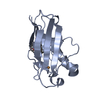
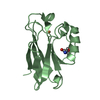
- PDB-1rkr: CRYSTAL STRUCTURE OF AZURIN-I FROM ALCALIGENES XYLOSOXIDANS NCIMB... -
+ Open data

- Basic information
| Entry | Database: PDB / ID: 1rkr | ||||||
|---|---|---|---|---|---|---|---|
| Title | CRYSTAL STRUCTURE OF AZURIN-I FROM ALCALIGENES XYLOSOXIDANS NCIMB 11015 | ||||||
 Components Components | AZURIN-I | ||||||
 Keywords Keywords | ELECTRON TRANSPORT | ||||||
| Function / homology |  Function and homology information Function and homology information | ||||||
| Biological species |  Achromobacter xylosoxidans (bacteria) Achromobacter xylosoxidans (bacteria) | ||||||
| Method | X-RAY DIFFRACTION / MOLECULAR REPLACEMENT / Resolution: 2.45 Å | ||||||
 Authors Authors | Li, C. / Inoue, T. / Gotowda, M. / Suzuki, S. / Yamaguchi, K. / Kataoka, K. / Kai, Y. | ||||||
 Citation Citation | Journal: Acta Crystallogr.,Sect.D / Year: 1998 Title: Structure of azurin I from the denitrifying bacterium Alcaligenes xylosoxidans NCIMB 11015 at 2.45 A resolution. Authors: Li, C. / Inoue, T. / Gotowda, M. / Suzuki, S. / Yamaguchi, K. / Kunishige, K. / Kai, Y. #1: Journal: Chem.Lett. / Year: 1995Title: Isolation and Characterization of Two Distinct Azurins from Alcaligenes Xylosoxidans Subsp. Xylosoxidans Ncib11015 or Gifu1051 Authors: Yamaguchi, K. / Deligeer / Nakamura, N. / Shidara, S. / Iwasaki, H. / Suzuki, S. #2: Journal: J.Biochem.(Tokyo) / Year: 1994Title: Structure of Azurin from Achromobacter Xylosoxidans Ncib11015 at 2.5 A Resolution Authors: Inoue, T. / Shibata, N. / Nakanishi, H. / Koyama, S. / Ishii, H. / Kai, Y. / Harada, S. / Kasai, N. / Ohshiro, Y. / Suzuki, S. / Kohzuma, T. / Yamaguchi, K. / Shidara, S. / Iwasaki, H. | ||||||
| History |
|
- Structure visualization
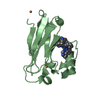
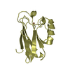
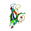
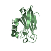
| Structure viewer | Molecule: MolmilJmol/JSmol |
|---|
- Downloads & links
-Download
| PDBx/mmCIF format | 1rkr.cif.gz | 108.2 KB | Display | PDBx/mmCIF format |
|---|---|---|---|---|
| PDB format | pdb1rkr.ent.gz | 84.7 KB | Display | PDB format |
| PDBx/mmJSON format | 1rkr.json.gz | Tree view | PDBx/mmJSON format | |
| Others |  Other downloads Other downloads |
-Validation report
| Arichive directory | https://data.pdbj.org/pub/pdb/validation_reports/rk/1rkrftp://data.pdbj.org/pub/pdb/validation_reports/rk/1rkr | HTTPS FTP |
|---|
-Related structure data
| Similar structure data |
|---|


-Links
 PDBj
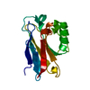
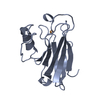
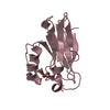
PDBj- Assembly
| Deposited unit | 
| ||||||||
|---|---|---|---|---|---|---|---|---|---|
| 1 | 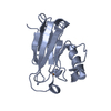
| ||||||||
| 2 | 
| ||||||||
| 3 | 
| ||||||||
| 4 | 
| ||||||||
| 5 | 
| ||||||||
| Unit cell |
|
-Components
| #1: Protein | Mass: 13904.841 Da / Num. of mol.: 4 / Source method: isolated from a natural source / Source: (natural) Achromobacter xylosoxidans (bacteria) / Strain: NCIMB 11015 / References: UniProt: P56547#2: Chemical | ChemComp-CU / Copper  Mass: 63.546 Da / Num. of mol.: 4 / Source method: obtained synthetically / Formula: Cu Mass: 63.546 Da / Num. of mol.: 4 / Source method: obtained synthetically / Formula: Cu#3: Water | ChemComp-HOH / | Water Mass: 18.015 Da / Num. of mol.: 81 / Source method: isolated from a natural source / Formula: H2O Mass: 18.015 Da / Num. of mol.: 81 / Source method: isolated from a natural source / Formula: H2O |
|---|
-Experimental details
-Experiment
| Experiment | Method: X-RAY DIFFRACTION / Number of used crystals: 1 |
|---|
- Sample preparation
| Crystal | Density Matthews: 2.35 Å3/Da / Density % sol: 48 % | ||||||||||||||||||||||||||||||
|---|---|---|---|---|---|---|---|---|---|---|---|---|---|---|---|---|---|---|---|---|---|---|---|---|---|---|---|---|---|---|---|
| Crystal grow | Method: vapor diffusion, hanging drop / pH: 8 Details: HANGING-DROP VAPOR DIFFUSION METHOD, pH 8.0, vapor diffusion - hanging drop | ||||||||||||||||||||||||||||||
| Crystal grow | *PLUS Method: vapor diffusion, hanging drop | ||||||||||||||||||||||||||||||
| Components of the solutions | *PLUS
|
-Data collection
| Diffraction | Mean temperature: 298 K |
|---|---|
| Diffraction source | Source: ROTATING ANODE / Type: RIGAKU RUH3R / Wavelength: 1.5418 |
| Detector | Type: RIGAKU / Detector: IMAGE PLATE / Date: Feb 20, 1996 |
| Radiation | Monochromator: GRAPHITE(002) / Monochromatic (M) / Laue (L): M / Scattering type: x-ray |
| Radiation wavelength | Wavelength: 1.5418 Å / Relative weight: 1 |
| Reflection | Resolution: 2.45→50 Å / Num. obs: 11462 / % possible obs: 90.5 % / Observed criterion σ(I): 1 / Redundancy: 1.7 % / Rmerge(I) obs: 0.069 / Net I/σ(I): 11.7 |
| Reflection shell | Resolution: 2.45→2.5 Å / Rmerge(I) obs: 0.171 / Mean I/σ(I) obs: 3.47 / % possible all: 82.3 |
| Reflection | *PLUS Num. measured all: 19618 |
| Reflection shell | *PLUS % possible obs: 82.3 % / Num. unique obs: 904 / Num. measured obs: 1087 |
- Processing
| Software |
| ||||||||||||||||||||||||||||||||||||||||||||||||||||||||||||
|---|---|---|---|---|---|---|---|---|---|---|---|---|---|---|---|---|---|---|---|---|---|---|---|---|---|---|---|---|---|---|---|---|---|---|---|---|---|---|---|---|---|---|---|---|---|---|---|---|---|---|---|---|---|---|---|---|---|---|---|---|---|
| Refinement | Method to determine structure: MOLECULAR REPLACEMENT Starting model: AZURIN-II FROM THE SAME BACTERIUM Resolution: 2.45→8 Å / Data cutoff low absF: 0 / σ(F): 2
| ||||||||||||||||||||||||||||||||||||||||||||||||||||||||||||
| Refine analyze | Luzzati coordinate error obs: 0.25 Å | ||||||||||||||||||||||||||||||||||||||||||||||||||||||||||||
| Refinement step | Cycle: LAST / Resolution: 2.45→8 Å
| ||||||||||||||||||||||||||||||||||||||||||||||||||||||||||||
| Refine LS restraints |
| ||||||||||||||||||||||||||||||||||||||||||||||||||||||||||||
| Software | *PLUS Name: X-PLOR / Version: 3 / Classification: refinement | ||||||||||||||||||||||||||||||||||||||||||||||||||||||||||||
| Refinement | *PLUS | ||||||||||||||||||||||||||||||||||||||||||||||||||||||||||||
| Solvent computation | *PLUS | ||||||||||||||||||||||||||||||||||||||||||||||||||||||||||||
| Displacement parameters | *PLUS | ||||||||||||||||||||||||||||||||||||||||||||||||||||||||||||
| Refine LS restraints | *PLUS
|
