 Movie
Movie Controller

[English] 日本語
 Yorodumi
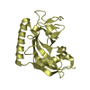
- PDB-6nm4: Crystal structure of SAM-bound PRDM9 in complex with MRK-740 inhibitor -
+ Open data

- Basic information
| Entry | Database: PDB / ID: 6nm4 | ||||||
|---|---|---|---|---|---|---|---|
| Title | Crystal structure of SAM-bound PRDM9 in complex with MRK-740 inhibitor | ||||||
 Components Components | Histone-lysine N-methyltransferase PRDM9 | ||||||
 Keywords Keywords | GENE REGULATION/INHIBITOR / PR SET domain / Lysine Methyltransferase / Inhibitor /  Structural Genomics / Structural Genomics Consortium / SGC / GENE REGULATION-INHIBITOR complex Structural Genomics / Structural Genomics Consortium / SGC / GENE REGULATION-INHIBITOR complex | ||||||
| Function / homology |  Function and homology informationrecombination hotspot binding / positive regulation of reciprocal meiotic recombination / positive regulation of fertilization / [histone H3]-lysine9 N-trimethyltransferase / male gamete generation / histone H4K20me methyltransferase activity / meiotic gene conversion / [histone H4]-N-methyl-L-lysine20 N-methyltransferase / histone H4K20 monomethyltransferase activity / histone H3K9 trimethyltransferase activity ...recombination hotspot binding / positive regulation of reciprocal meiotic recombination / positive regulation of fertilization / [histone H3]-lysine9 N-trimethyltransferase / male gamete generation / histone H4K20me methyltransferase activity / meiotic gene conversion / [histone H4]-N-methyl-L-lysine20 N-methyltransferase / histone H4K20 monomethyltransferase activity / histone H3K9 trimethyltransferase activity / [histone H4]-lysine20 N-methyltransferase / [histone H3]-lysine36 N-trimethyltransferase / female gamete generation / histone H3K36 trimethyltransferase activity / [histone H3]-lysine4 N-trimethyltransferase / homologous chromosome pairing at meiosis / double-strand break repair involved in meiotic recombination / histone H3K4 trimethyltransferase activity / histone H3K36 methyltransferase activity / histone H3K4 methyltransferase activity / Transferases; Transferring one-carbon groups; Methyltransferases / PKMTs methylate histone lysines / Meiotic recombination / chromosome / methylation / regulation of gene expression / transcription cis-regulatory region binding / regulation of DNA-templated transcription / negative regulation of apoptotic process / protein homodimerization activity / zinc ion binding / nucleoplasm / nucleus Function and homology informationrecombination hotspot binding / positive regulation of reciprocal meiotic recombination / positive regulation of fertilization / [histone H3]-lysine9 N-trimethyltransferase / male gamete generation / histone H4K20me methyltransferase activity / meiotic gene conversion / [histone H4]-N-methyl-L-lysine20 N-methyltransferase / histone H4K20 monomethyltransferase activity / histone H3K9 trimethyltransferase activity ...recombination hotspot binding / positive regulation of reciprocal meiotic recombination / positive regulation of fertilization / [histone H3]-lysine9 N-trimethyltransferase / male gamete generation / histone H4K20me methyltransferase activity / meiotic gene conversion / [histone H4]-N-methyl-L-lysine20 N-methyltransferase / histone H4K20 monomethyltransferase activity / histone H3K9 trimethyltransferase activity / [histone H4]-lysine20 N-methyltransferase / [histone H3]-lysine36 N-trimethyltransferase / female gamete generation / histone H3K36 trimethyltransferase activity / [histone H3]-lysine4 N-trimethyltransferase / homologous chromosome pairing at meiosis / double-strand break repair involved in meiotic recombination / histone H3K4 trimethyltransferase activity / histone H3K36 methyltransferase activity / histone H3K4 methyltransferase activity / Transferases; Transferring one-carbon groups; Methyltransferases / PKMTs methylate histone lysines / Meiotic recombination / chromosome / methylation / regulation of gene expression / transcription cis-regulatory region binding / regulation of DNA-templated transcription / negative regulation of apoptotic process / protein homodimerization activity / zinc ion binding / nucleoplasm / nucleusSimilarity search - Function | ||||||
| Biological species |  Homo sapiens (human) Homo sapiens (human) | ||||||
| Method | X-RAY DIFFRACTION / SYNCHROTRON / MOLECULAR REPLACEMENT / molecular replacement / Resolution: 2.58 Å | ||||||
 Authors Authors | Ivanochko, D. / Halabelian, L. / Fischer, C. / Sanders, J.M. / Kattar, S.D. / Brown, P.J. / Edwards, A.M. / Bountra, C. / Arrowsmith, C.H. / Structural Genomics Consortium (SGC) | ||||||
 Citation Citation | Journal: Nat Commun / Year: 2019 Title: Discovery of a chemical probe for PRDM9. Authors: Allali-Hassani, A. / Szewczyk, M.M. / Ivanochko, D. / Organ, S.L. / Bok, J. / Ho, J.S.Y. / Gay, F.P.H. / Li, F. / Blazer, L. / Eram, M.S. / Halabelian, L. / Dilworth, D. / Luciani, G.M. / ...Authors: Allali-Hassani, A. / Szewczyk, M.M. / Ivanochko, D. / Organ, S.L. / Bok, J. / Ho, J.S.Y. / Gay, F.P.H. / Li, F. / Blazer, L. / Eram, M.S. / Halabelian, L. / Dilworth, D. / Luciani, G.M. / Lima-Fernandes, E. / Wu, Q. / Loppnau, P. / Palmer, N. / Talib, S.Z.A. / Brown, P.J. / Schapira, M. / Kaldis, P. / O'Hagan, R.C. / Guccione, E. / Barsyte-Lovejoy, D. / Arrowsmith, C.H. / Sanders, J.M. / Kattar, S.D. / Bennett, D.J. / Nicholson, B. / Vedadi, M. | ||||||
| History |
|
- Structure visualization
| Structure viewer | Molecule: MolmilJmol/JSmol |
|---|
- Downloads & links
-Download
| PDBx/mmCIF format | 6nm4.cif.gz | 172.6 KB | Display | PDBx/mmCIF format |
|---|---|---|---|---|
| PDB format | pdb6nm4.ent.gz | 134.5 KB | Display | PDB format |
| PDBx/mmJSON format | 6nm4.json.gz | Tree view | PDBx/mmJSON format | |
| Others |  Other downloads Other downloads |
-Validation report
| Arichive directory | https://data.pdbj.org/pub/pdb/validation_reports/nm/6nm4ftp://data.pdbj.org/pub/pdb/validation_reports/nm/6nm4 | HTTPS FTP |
|---|
-Related structure data
| Related structure data |  4ijdS S: Starting model for refinement |
|---|---|
| Similar structure data |


-Links
 PDBj
PDBj


- Assembly
| Deposited unit | 
| ||||||||||||||||||
|---|---|---|---|---|---|---|---|---|---|---|---|---|---|---|---|---|---|---|---|
| 1 | 
| ||||||||||||||||||
| 2 | 
| ||||||||||||||||||
| Unit cell |
| ||||||||||||||||||
| Noncrystallographic symmetry (NCS) | NCS domain:
NCS domain segments: Component-ID: 0 / Ens-ID: 1 / Beg auth comp-ID: GLU / Beg label comp-ID: GLU / End auth comp-ID: LEU / End label comp-ID: LEU / Refine code: 0 / Auth seq-ID: 196 - 377 / Label seq-ID: 2 - 183
|
-Components
-Protein , 1 types, 2 molecules AB
| #1: Protein | Mass: 21802.352 Da / Num. of mol.: 2 / Fragment: PR-SET domain (UNP residues 195-385) Source method: isolated from a genetically manipulated source Source: (gene. exp.) Homo sapiens (human) / Gene: PRDM9, PFM6 / Production host:  Escherichia coli (E. coli) Escherichia coli (E. coli)References: UniProt: Q9NQV7, histone-lysine N-methyltransferase |
|---|
-Non-polymers , 5 types, 32 molecules 


| #2: Chemical |  Mass: 65.409 Da / Num. of mol.: 2 / Source method: obtained synthetically / Formula: Zn Mass: 65.409 Da / Num. of mol.: 2 / Source method: obtained synthetically / Formula: Zn#3: Chemical | S-Adenosyl methionine Mass: 398.437 Da / Num. of mol.: 2 / Source method: obtained synthetically / Formula: C15H22N6O5S Mass: 398.437 Da / Num. of mol.: 2 / Source method: obtained synthetically / Formula: C15H22N6O5S#4: Chemical |  Mass: 464.560 Da / Num. of mol.: 2 / Source method: obtained synthetically / Formula: C25H32N6O3 Mass: 464.560 Da / Num. of mol.: 2 / Source method: obtained synthetically / Formula: C25H32N6O3#5: Chemical |  Num. of mol.: 3 / Source method: obtained synthetically Num. of mol.: 3 / Source method: obtained synthetically#6: Water | ChemComp-HOH / | WaterMass: 18.015 Da / Num. of mol.: 23 / Source method: isolated from a natural source / Formula: H2O |
|---|
-Experimental details
-Experiment
| Experiment | Method: X-RAY DIFFRACTION / Number of used crystals: 1 |
|---|
- Sample preparation
| Crystal | Density Matthews: 2.38 Å3/Da / Density % sol: 48.3 % |
|---|---|
| Crystal grow | Temperature: 291 K / Method: vapor diffusion, sitting drop Details: 0.1 M Bis-Tris, pH 6.0, 0.2 M ammonium acetate, 24.5% PEG3350 |
-Data collection
| Diffraction | Mean temperature: 100 K / Serial crystal experiment: N | ||||||||||||||||||||||||
|---|---|---|---|---|---|---|---|---|---|---|---|---|---|---|---|---|---|---|---|---|---|---|---|---|---|
| Diffraction source | Source: SYNCHROTRON / Site: APS  / Beamline: 24-ID-E / Wavelength: 0.97918 Å / Beamline: 24-ID-E / Wavelength: 0.97918 Å | ||||||||||||||||||||||||
| Detector | Type: DECTRIS EIGER X 16M / Detector: PIXEL / Date: Feb 24, 2018 | ||||||||||||||||||||||||
| Radiation | Monochromator: Cryogenically-cooled single crystal Si(220) side bounce Protocol: SINGLE WAVELENGTH / Monochromatic (M) / Laue (L): M / Scattering type: x-ray | ||||||||||||||||||||||||
| Radiation wavelength | Wavelength: 0.97918 Å / Relative weight: 1 | ||||||||||||||||||||||||
| Reflection | Resolution: 2.58→39.88 Å / Num. obs: 13290 / % possible obs: 99.6 % / Redundancy: 7.3 % / CC1/2: 0.997 / Rmerge(I) obs: 0.129 / Rpim(I) all: 0.051 / Rrim(I) all: 0.139 / Net I/σ(I): 10.7 | ||||||||||||||||||||||||
| Reflection shell | Diffraction-ID: 1
|
-Phasing
| Phasing | Method: molecular replacement | |||||||||
|---|---|---|---|---|---|---|---|---|---|---|
| Phasing MR | Model details: Phaser MODE: MR_AUTO
|
- Processing
| Software |
| |||||||||||||||||||||||||||||||||||||||||||||||||||||||||||||||||||||||||||
|---|---|---|---|---|---|---|---|---|---|---|---|---|---|---|---|---|---|---|---|---|---|---|---|---|---|---|---|---|---|---|---|---|---|---|---|---|---|---|---|---|---|---|---|---|---|---|---|---|---|---|---|---|---|---|---|---|---|---|---|---|---|---|---|---|---|---|---|---|---|---|---|---|---|---|---|---|
| Refinement | Method to determine structure: MOLECULAR REPLACEMENT Starting model: PDB entry 4IJD Resolution: 2.58→39.88 Å / Cor.coef. Fo:Fc: 0.95 / Cor.coef. Fo:Fc free: 0.928 / SU B: 29.499 / SU ML: 0.285 / Cross valid method: THROUGHOUT / σ(F): 0 / ESU R: 1.759 / ESU R Free: 0.336 Details: HYDROGENS HAVE BEEN ADDED IN THE RIDING POSITIONS U VALUES : WITH TLS ADDED
| |||||||||||||||||||||||||||||||||||||||||||||||||||||||||||||||||||||||||||
| Solvent computation | Ion probe radii: 0.8 Å / Shrinkage radii: 0.8 Å / VDW probe radii: 1.2 Å | |||||||||||||||||||||||||||||||||||||||||||||||||||||||||||||||||||||||||||
| Displacement parameters | Biso max: 120.05 Å2 / Biso mean: 61.466 Å2 / Biso min: 30.37 Å2
| |||||||||||||||||||||||||||||||||||||||||||||||||||||||||||||||||||||||||||
| Refinement step | Cycle: final / Resolution: 2.58→39.88 Å
| |||||||||||||||||||||||||||||||||||||||||||||||||||||||||||||||||||||||||||
| Refine LS restraints |
| |||||||||||||||||||||||||||||||||||||||||||||||||||||||||||||||||||||||||||
| Refine LS restraints NCS | Ens-ID: 1 / Number: 5587 / Refine-ID: X-RAY DIFFRACTION / Type: interatomic distance / Rms dev position: 0.1 Å / Weight position: 0.05
| |||||||||||||||||||||||||||||||||||||||||||||||||||||||||||||||||||||||||||
| LS refinement shell | Resolution: 2.58→2.647 Å / Rfactor Rfree error: 0 / Total num. of bins used: 20
| |||||||||||||||||||||||||||||||||||||||||||||||||||||||||||||||||||||||||||
| Refinement TLS params. | Method: refined / Refine-ID: X-RAY DIFFRACTION
| |||||||||||||||||||||||||||||||||||||||||||||||||||||||||||||||||||||||||||
| Refinement TLS group |
|
