 Movie
Movie Controller


+ Open data

- Basic information
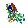
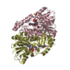
| Entry | Database: PDB / ID: 6m7a | |||||||||
|---|---|---|---|---|---|---|---|---|---|---|
| Title | Structure of REV7-R124A complexed with SHLD3(28-73) | |||||||||
 Components Components |
| |||||||||
 Keywords Keywords |  REPLICATION / REV7 / shieldin / SHLD3 / DNA damage REPLICATION / REV7 / shieldin / SHLD3 / DNA damage | |||||||||
| Function / homology |  Function and homology information Function and homology informationsomatic diversification of immunoglobulins involved in immune response / DNA damage response, signal transduction resulting in transcription / telomere maintenance in response to DNA damage / negative regulation of transcription regulatory region DNA binding / zeta DNA polymerase complex / positive regulation of isotype switching / positive regulation of extracellular matrix assembly / negative regulation of epithelial to mesenchymal transition / negative regulation of transcription by competitive promoter binding / negative regulation of cell-cell adhesion mediated by cadherin ...somatic diversification of immunoglobulins involved in immune response / DNA damage response, signal transduction resulting in transcription / telomere maintenance in response to DNA damage / negative regulation of transcription regulatory region DNA binding / zeta DNA polymerase complex / positive regulation of isotype switching / positive regulation of extracellular matrix assembly / negative regulation of epithelial to mesenchymal transition / negative regulation of transcription by competitive promoter binding / negative regulation of cell-cell adhesion mediated by cadherin / JUN kinase binding / negative regulation of ubiquitin protein ligase activity / negative regulation of double-strand break repair via homologous recombination / mitotic spindle assembly checkpoint signaling / positive regulation of double-strand break repair via nonhomologous end joining / positive regulation of epithelial to mesenchymal transition / error-prone translesion synthesis / Translesion synthesis by REV1 / Translesion synthesis by POLK / Translesion synthesis by POLI / actin filament organization / regulation of cell growth / negative regulation of canonical Wnt signaling pathway / negative regulation of DNA-binding transcription factor activity / negative regulation of protein catabolic process / spindle / double-strand break repair / positive regulation of peptidyl-serine phosphorylation / site of double-strand break / chromosome / RNA polymerase II-specific DNA-binding transcription factor binding / cell division / DNA repair / chromatin / nucleolus / positive regulation of DNA-templated transcription / negative regulation of transcription by RNA polymerase II / nucleoplasm / nucleus / cytoplasmSimilarity search - Function | |||||||||
| Biological species |  Homo sapiens (human) Homo sapiens (human) | |||||||||
| Method | X-RAY DIFFRACTION / SYNCHROTRON / MOLECULAR REPLACEMENT / Resolution: 1.9 Å | |||||||||
 Authors Authors | Ma, Y.Z. / Li, Y. / Wu, B.X. / Huang, H.D. | |||||||||
| Funding support |  China, 2items China, 2items
| |||||||||
 Citation Citation | Journal: To Be Published Title: Structure of REV7-R124A complexed with SHLD3(28-73) Authors: Ma, Y.Z. / Li, Y. / Wu, B.X. / Huang, H.D. | |||||||||
| History |
|
- Structure visualization
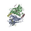
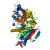
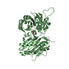
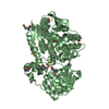
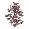
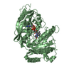
| Structure viewer | Molecule: MolmilJmol/JSmol |
|---|
- Downloads & links
-Download
| PDBx/mmCIF format | 6m7a.cif.gz | 125.9 KB | Display | PDBx/mmCIF format |
|---|---|---|---|---|
| PDB format | pdb6m7a.ent.gz | 88.5 KB | Display | PDB format |
| PDBx/mmJSON format | 6m7a.json.gz | Tree view | PDBx/mmJSON format | |
| Others |  Other downloads Other downloads |
-Validation report
| Arichive directory | https://data.pdbj.org/pub/pdb/validation_reports/m7/6m7aftp://data.pdbj.org/pub/pdb/validation_reports/m7/6m7a | HTTPS FTP |
|---|
-Related structure data
| Related structure data | 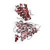 3vu7S S: Starting model for refinement |
|---|---|
| Similar structure data |

-Links
 PDBj
PDBj


- Assembly
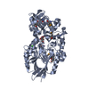
| Deposited unit | 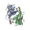
| ||||||||||||
|---|---|---|---|---|---|---|---|---|---|---|---|---|---|
| 1 |
| ||||||||||||
| Unit cell |
|
-Components
| #1: Protein | Mass: 24281.350 Da / Num. of mol.: 2 / Mutation: R124A Source method: isolated from a genetically manipulated source Source: (gene. exp.) Homo sapiens (human) / Gene: MAD2L2, MAD2B, REV7 / Production host:  Escherichia coli (E. coli) / References: UniProt: Q9UI95 Escherichia coli (E. coli) / References: UniProt: Q9UI95#2: Protein/peptide | Mass: 5381.123 Da / Num. of mol.: 2 Source method: isolated from a genetically manipulated source Source: (gene. exp.) Homo sapiens (human) / Gene: SHLD3, FLJ26957, RINN1 / Production host: Escherichia coli (E. coli) / References: UniProt: Q6ZNX1#3: Water | ChemComp-HOH / | Water Mass: 18.015 Da / Num. of mol.: 381 / Source method: isolated from a natural source / Formula: H2O Mass: 18.015 Da / Num. of mol.: 381 / Source method: isolated from a natural source / Formula: H2O |
|---|
-Experimental details
-Experiment
| Experiment | Method: X-RAY DIFFRACTION / Number of used crystals: 1 |
|---|
- Sample preparation
| Crystal | Density Matthews: 1.93 Å3/Da / Density % sol: 36.29 % |
|---|---|
| Crystal grow | Temperature: 293 K / Method: vapor diffusion, sitting drop Details: 0.2M NaCl Sodium Chloride; 0.1 M Na/K phosphate 6.2; 20% PEG1000 |
-Data collection
| Diffraction | Mean temperature: 100 K / Serial crystal experiment: N |
|---|---|
| Diffraction source | Source: SYNCHROTRON / Site: SSRF / Beamline: BL19U1 / Wavelength: 0.9785 Å |
| Detector | Type: DECTRIS PILATUS3 S 6M / Detector: PIXEL / Date: Jan 7, 2019 |
| Radiation | Protocol: SINGLE WAVELENGTH / Monochromatic (M) / Laue (L): M / Scattering type: x-ray |
| Radiation wavelength | Wavelength: 0.9785 Å / Relative weight: 1 |
| Reflection | Resolution: 1.9→40 Å / Num. obs: 37019 / % possible obs: 100 % / Redundancy: 11.2 % / CC1/2: 0.433 / Net I/σ(I): 2.4 |
| Reflection shell | Resolution: 1.9→1.97 Å / Num. unique obs: 3640 / CC1/2: 0.45 |
- Processing
| Software |
| ||||||||||||||||||||||||
|---|---|---|---|---|---|---|---|---|---|---|---|---|---|---|---|---|---|---|---|---|---|---|---|---|---|
| Refinement | Method to determine structure: MOLECULAR REPLACEMENT Starting model: 3VU7 Resolution: 1.9→40 Å / Cross valid method: FREE R-VALUE
| ||||||||||||||||||||||||
| Displacement parameters | Biso mean: 27.37 Å2 | ||||||||||||||||||||||||
| Refinement step | Cycle: LAST / Resolution: 1.9→40 Å
| ||||||||||||||||||||||||
| Refine LS restraints |
| ||||||||||||||||||||||||
| LS refinement shell | Resolution: 1.8969→1.9482 Å
|
