 Movie
Movie Controller

[English] 日本語
 Yorodumi
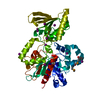
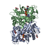
- PDB-2gp4: Structure of [FeS]cluster-free Apo Form of 6-Phosphogluconate Deh... -
+ Open data

- Basic information
| Entry | Database: PDB / ID: 2gp4 | ||||||
|---|---|---|---|---|---|---|---|
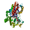
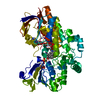
| Title | Structure of [FeS]cluster-free Apo Form of 6-Phosphogluconate Dehydratase from Shewanella oneidensis | ||||||
 Components Components | 6-phosphogluconate dehydratase | ||||||
 Keywords Keywords |  LYASE / N-terminal domain largely alpha-helical / C-terminal domain mainly beta-sheet (trefoil-like) / Structural Genomics / PSI / Protein Structure Initiative / Southeast Collaboratory for Structural Genomics / SECSG LYASE / N-terminal domain largely alpha-helical / C-terminal domain mainly beta-sheet (trefoil-like) / Structural Genomics / PSI / Protein Structure Initiative / Southeast Collaboratory for Structural Genomics / SECSG | ||||||
| Function / homology |  Function and homology informationphosphogluconate dehydratase / phosphogluconate dehydratase activity / Entner-Doudoroff pathway through 6-phosphogluconate / D-gluconate catabolic process / hydro-lyase activity / 4 iron, 4 sulfur cluster binding / metal ion binding / cytosol Function and homology informationphosphogluconate dehydratase / phosphogluconate dehydratase activity / Entner-Doudoroff pathway through 6-phosphogluconate / D-gluconate catabolic process / hydro-lyase activity / 4 iron, 4 sulfur cluster binding / metal ion binding / cytosolSimilarity search - Function | ||||||
| Biological species |  Shewanella oneidensis (bacteria) Shewanella oneidensis (bacteria) | ||||||
| Method | X-RAY DIFFRACTION / SYNCHROTRON / SAD / Resolution: 2.49 Å | ||||||
 Authors Authors | Schormann, N. / Symersky, J. / Southeast Collaboratory for Structural Genomics (SECSG) | ||||||
 Citation Citation | Journal: To be Published Title: Structure of [FeS]cluster-free Apo Form of 6-Phosphogluconate Dehydratase from Shewanella oneidensis Authors: Schormann, N. / Symersky, J. / Karpova, E. / Zhang, Y. / Lu, S. / Qiu, S. / Bunzel, R. / Luan, C.-H. / Huang, W. / Luo, M. / Tsao, J. / Johnson, D. / Carson, M. / Zhou, Q. / Luo, D. / Gray, ...Authors: Schormann, N. / Symersky, J. / Karpova, E. / Zhang, Y. / Lu, S. / Qiu, S. / Bunzel, R. / Luan, C.-H. / Huang, W. / Luo, M. / Tsao, J. / Johnson, D. / Carson, M. / Zhou, Q. / Luo, D. / Gray, R. / Cao, Z. / An, J. / Arabshahi, A. / Li, S. / Stinnett, M. / McKinstry, A. / Lin, G. / Shang, Q. / Chen, Y. / DeLucas, L. | ||||||
| History |
|
- Structure visualization
| Structure viewer | Molecule: MolmilJmol/JSmol |
|---|
- Downloads & links
-Download
| PDBx/mmCIF format | 2gp4.cif.gz | 209.1 KB | Display | PDBx/mmCIF format |
|---|---|---|---|---|
| PDB format | pdb2gp4.ent.gz | 171.9 KB | Display | PDB format |
| PDBx/mmJSON format | 2gp4.json.gz | Tree view | PDBx/mmJSON format | |
| Others |  Other downloads Other downloads |
-Validation report
| Arichive directory | https://data.pdbj.org/pub/pdb/validation_reports/gp/2gp4ftp://data.pdbj.org/pub/pdb/validation_reports/gp/2gp4 | HTTPS FTP |
|---|
-Related structure data
| Similar structure data | |
|---|---|
| Other databases |

-Links
 PDBj
PDBj- Assembly
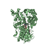
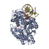
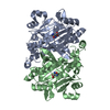
| Deposited unit | 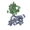
| ||||||||||||||||||
|---|---|---|---|---|---|---|---|---|---|---|---|---|---|---|---|---|---|---|---|
| 1 | 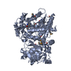
| ||||||||||||||||||
| 2 | 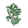
| ||||||||||||||||||
| 3 | 
| ||||||||||||||||||
| Unit cell |
| ||||||||||||||||||
| Noncrystallographic symmetry (NCS) | NCS domain:
NCS domain segments: Component-ID: 1 / Ens-ID: 1 / Beg label comp-ID: HIS / End label comp-ID: TYR / Refine code: 4 / Auth seq-ID: 0 - 608 / Label seq-ID: 20 - 628
| ||||||||||||||||||
| Details | The biological assembly is likely a monomer. The asymmetric unit contains two monomers related by a non-crystallographic 2-fold. |
-Components
| #1: Protein | Mass: 67696.336 Da / Num. of mol.: 2 Source method: isolated from a genetically manipulated source Source: (gene. exp.) Shewanella oneidensis (bacteria) / Strain: MR-1 / Gene: edd / Plasmid: pET28b / Production host: Escherichia coli (E. coli) / Strain (production host): B834References: GenBank: 24348501, UniProt: Q8EEA0*PLUS, phosphogluconate dehydratase#2: Water | ChemComp-HOH / | Water Mass: 18.015 Da / Num. of mol.: 553 / Source method: isolated from a natural source / Formula: H2O Mass: 18.015 Da / Num. of mol.: 553 / Source method: isolated from a natural source / Formula: H2O |
|---|
-Experimental details
-Experiment
| Experiment | Method: X-RAY DIFFRACTION / Number of used crystals: 1 |
|---|
- Sample preparation
| Crystal | Density Matthews: 2.15 Å3/Da / Density % sol: 42.76 % |
|---|---|
| Crystal grow | Temperature: 295 K / Method: vapor diffusion, hanging drop / pH: 5.6 Details: 5% PEG400, 50mM cacodylate, 3mM DTT, pH 5.6, VAPOR DIFFUSION, HANGING DROP, temperature 295K |
-Data collection
| Diffraction | Mean temperature: 100 K |
|---|---|
| Diffraction source | Source: SYNCHROTRON / Site: APS  / Beamline: 22-ID / Wavelength: 0.9792 Å / Beamline: 22-ID / Wavelength: 0.9792 Å |
| Detector | Type: MARMOSAIC 225 mm CCD / Detector: CCD / Date: Dec 8, 2004 |
| Radiation | Monochromator: Graphite / Protocol: SINGLE WAVELENGTH / Monochromatic (M) / Laue (L): M / Scattering type: x-ray |
| Radiation wavelength | Wavelength: 0.9792 Å / Relative weight: 1 |
| Reflection | Resolution: 2.49→50 Å / Num. all: 41534 / Num. obs: 41534 / % possible obs: 99.8 % / Observed criterion σ(F): 1 / Observed criterion σ(I): 1 / Redundancy: 9.8 % / Rmerge(I) obs: 0.08 / Rsym value: 0.08 / Net I/σ(I): 15.1 |
| Reflection shell | Resolution: 2.49→2.59 Å / Redundancy: 8.6 % / Rmerge(I) obs: 0.206 / Num. unique all: 4092 / Rsym value: 0.206 / % possible all: 99.8 |
- Processing
| Software |
| ||||||||||||||||||||||||||||||||||||||||||||||||||||||||||||||||||||||||||||||||||||||||||
|---|---|---|---|---|---|---|---|---|---|---|---|---|---|---|---|---|---|---|---|---|---|---|---|---|---|---|---|---|---|---|---|---|---|---|---|---|---|---|---|---|---|---|---|---|---|---|---|---|---|---|---|---|---|---|---|---|---|---|---|---|---|---|---|---|---|---|---|---|---|---|---|---|---|---|---|---|---|---|---|---|---|---|---|---|---|---|---|---|---|---|---|
| Refinement | Method to determine structure: SAD / Resolution: 2.49→48.68 Å / Cor.coef. Fo:Fc: 0.905 / Cor.coef. Fo:Fc free: 0.845 / SU B: 17.206 / SU ML: 0.214 / TLS residual ADP flag: LIKELY RESIDUAL / Isotropic thermal model: Isotropic / Cross valid method: THROUGHOUT / σ(F): 0 / ESU R: 0.629 / ESU R Free: 0.335 / Stereochemistry target values: MAXIMUM LIKELIHOOD Details: The N-terminal His-tag (-19 to -1) is not visible in the electron density except for His (0). The inactive apo protein (no substrate) is cluster-free. The N-terminal domain, which normally ...Details: The N-terminal His-tag (-19 to -1) is not visible in the electron density except for His (0). The inactive apo protein (no substrate) is cluster-free. The N-terminal domain, which normally contains the active site with a [4Fe-4S] cluster and a site for substrate-binding is largely disordered. Residues 34-89, 93-95, 106, 110-11, 181-199 and 226-230 are not visible in the electron density in chain a. in chain b residues 34-92, 106-107, 110-111, 155 and 181-230 are disordered and not visible in the electron density. In addition, side chains of residues 95, 97 and 98 in chain b are truncated because of missing electron density.
| ||||||||||||||||||||||||||||||||||||||||||||||||||||||||||||||||||||||||||||||||||||||||||
| Solvent computation | Ion probe radii: 0.8 Å / Shrinkage radii: 0.8 Å / VDW probe radii: 1.4 Å / Solvent model: MASK | ||||||||||||||||||||||||||||||||||||||||||||||||||||||||||||||||||||||||||||||||||||||||||
| Displacement parameters | Biso mean: 29.211 Å2
| ||||||||||||||||||||||||||||||||||||||||||||||||||||||||||||||||||||||||||||||||||||||||||
| Refinement step | Cycle: LAST / Resolution: 2.49→48.68 Å
| ||||||||||||||||||||||||||||||||||||||||||||||||||||||||||||||||||||||||||||||||||||||||||
| Refine LS restraints |
| ||||||||||||||||||||||||||||||||||||||||||||||||||||||||||||||||||||||||||||||||||||||||||
| Refine LS restraints NCS | Dom-ID: 1 / Auth asym-ID: A / Ens-ID: 1 / Number: 3642 / Refine-ID: X-RAY DIFFRACTION
| ||||||||||||||||||||||||||||||||||||||||||||||||||||||||||||||||||||||||||||||||||||||||||
| LS refinement shell | Resolution: 2.491→2.556 Å / Total num. of bins used: 20
| ||||||||||||||||||||||||||||||||||||||||||||||||||||||||||||||||||||||||||||||||||||||||||
| Refinement TLS params. | Method: refined / Refine-ID: X-RAY DIFFRACTION
| ||||||||||||||||||||||||||||||||||||||||||||||||||||||||||||||||||||||||||||||||||||||||||
| Refinement TLS group |
|
