 Movie
Movie Controller


+ Open data

- Basic information
| Entry | Database: PDB / ID: 3vu7 | ||||||
|---|---|---|---|---|---|---|---|
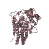
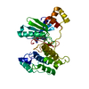
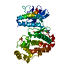
| Title | Crystal structure of REV1-REV7-REV3 ternary complex | ||||||
 Components Components |
| ||||||
 Keywords Keywords |  REPLICATION / DNA polymerase / DNA replication / Translesion DNA synthesis / DNA damage tolerance / DNA repair REPLICATION / DNA polymerase / DNA replication / Translesion DNA synthesis / DNA damage tolerance / DNA repair | ||||||
| Function / homology |  Function and homology information Function and homology informationsomatic diversification of immunoglobulins involved in immune response / DNA damage response, signal transduction resulting in transcription / deoxycytidyl transferase activity / telomere maintenance in response to DNA damage / negative regulation of transcription regulatory region DNA binding / zeta DNA polymerase complex / positive regulation of isotype switching / positive regulation of extracellular matrix assembly / negative regulation of transcription by competitive promoter binding / negative regulation of cell-cell adhesion mediated by cadherin ...somatic diversification of immunoglobulins involved in immune response / DNA damage response, signal transduction resulting in transcription / deoxycytidyl transferase activity / telomere maintenance in response to DNA damage / negative regulation of transcription regulatory region DNA binding / zeta DNA polymerase complex / positive regulation of isotype switching / positive regulation of extracellular matrix assembly / negative regulation of transcription by competitive promoter binding / negative regulation of cell-cell adhesion mediated by cadherin / negative regulation of epithelial to mesenchymal transition / JUN kinase binding / negative regulation of ubiquitin protein ligase activity / error-free translesion synthesis / mitotic spindle assembly checkpoint signaling / positive regulation of double-strand break repair via nonhomologous end joining / negative regulation of double-strand break repair via homologous recombination / error-prone translesion synthesis / positive regulation of epithelial to mesenchymal transition / response to UV / Translesion synthesis by REV1 / Translesion synthesis by POLK / Translesion synthesis by POLI / actin filament organization / regulation of cell growth / Termination of translesion DNA synthesis / double-strand break repair via homologous recombination / negative regulation of canonical Wnt signaling pathway / negative regulation of protein catabolic process / negative regulation of DNA-binding transcription factor activity / DNA-templated DNA replication / spindle / double-strand break repair / Transferases; Transferring phosphorus-containing groups; Nucleotidyltransferases / positive regulation of peptidyl-serine phosphorylation / site of double-strand break / chromosome / 4 iron, 4 sulfur cluster binding / RNA polymerase II-specific DNA-binding transcription factor binding / DNA replication / damaged DNA binding / DNA-directed DNA polymerase / DNA-directed DNA polymerase activity / cell division / nucleotide binding / chromatin / nucleolus / positive regulation of DNA-templated transcription / negative regulation of transcription by RNA polymerase II / DNA binding / nucleoplasm / metal ion binding / nucleus / cytoplasmSimilarity search - Function | ||||||
| Biological species |  Homo sapiens (human) Homo sapiens (human) | ||||||
| Method | X-RAY DIFFRACTION / SYNCHROTRON / MOLECULAR REPLACEMENT / Resolution: 2.8 Å | ||||||
 Authors Authors | Kikuchi, S. / Hara, K. / Shimizu, T. / Sato, M. / Hashimoto, H. | ||||||
 Citation Citation | Journal: J.Biol.Chem. / Year: 2012 Title: Structural basis of recruitment of DNA polymerase [zeta] by interaction between REV1 and REV7 proteins Authors: Kikuchi, S. / Hara, K. / Shimizu, T. / Sato, M. / Hashimoto, H. | ||||||
| History |
|
- Structure visualization
| Structure viewer | Molecule: MolmilJmol/JSmol |
|---|
- Downloads & links
-Download
| PDBx/mmCIF format | 3vu7.cif.gz | 143 KB | Display | PDBx/mmCIF format |
|---|---|---|---|---|
| PDB format | pdb3vu7.ent.gz | 112.2 KB | Display | PDB format |
| PDBx/mmJSON format | 3vu7.json.gz | Tree view | PDBx/mmJSON format | |
| Others |  Other downloads Other downloads |
-Validation report
| Arichive directory | https://data.pdbj.org/pub/pdb/validation_reports/vu/3vu7ftp://data.pdbj.org/pub/pdb/validation_reports/vu/3vu7 | HTTPS FTP |
|---|
-Related structure data
| Related structure data |  3abdS S: Starting model for refinement |
|---|---|
| Similar structure data |


-Links
 PDBj
PDBj


- Assembly
| Deposited unit | 
| ||||||||
|---|---|---|---|---|---|---|---|---|---|
| 1 |
| ||||||||
| Unit cell |
|
-Components
| #1: Protein | / Alpha integrin-binding protein 80 / AIBP80 / Rev1-like terminal deoxycytidyl transferase Mass: 14095.096 Da / Num. of mol.: 1 / Fragment: UNP residues 1140-1251 Source method: isolated from a genetically manipulated source Source: (gene. exp.) Homo sapiens (human) / Gene: REV1, REV1L / Production host:  Escherichia coli (E. coli) Escherichia coli (E. coli)References: UniProt: Q9UBZ9, Transferases; Transferring phosphorus-containing groups; Nucleotidyltransferases |
|---|---|
| #2: Protein | Mass: 26101.236 Da / Num. of mol.: 1 Source method: isolated from a genetically manipulated source Source: (gene. exp.) Homo sapiens (human) / Gene: MAD2L2, MAD2B, REV7 / Production host: Escherichia coli (E. coli) / References: UniProt: Q9UI95 |
| #3: Protein | Mass: 5632.319 Da / Num. of mol.: 1 / Fragment: UNP residues 1847-1898 / Mutation: R124A Source method: isolated from a genetically manipulated source Source: (gene. exp.) Homo sapiens (human) / Gene: REV3L, POLZ, REV3 / Production host: Escherichia coli (E. coli) / References: UniProt: O60673, DNA-directed DNA polymerase |
-Experimental details
-Experiment
| Experiment | Method: X-RAY DIFFRACTION |
|---|
- Sample preparation
| Crystal | Density Matthews: 2.19 Å3/Da / Density % sol: 43.8 % |
|---|
-Data collection
| Diffraction source | Source: SYNCHROTRON / Site: SPring-8  / Beamline: BL32XU / Wavelength: 1 Å / Beamline: BL32XU / Wavelength: 1 Å |
|---|---|
| Detector | Date: Jun 19, 2012 |
| Radiation | Protocol: SINGLE WAVELENGTH / Monochromatic (M) / Laue (L): M / Scattering type: x-ray |
| Radiation wavelength | Wavelength: 1 Å / Relative weight: 1 |
| Reflection | Resolution: 2.8→65.01 Å / Num. obs: 10306 |
- Processing
| Software | Name: REFMAC / Version: 5.6.0117 / Classification: refinement | ||||||||||||||||||||||||||||||||||||||||||||||||||||||||||||||||||||||||||||||||||||||||||||||||||||||||||||||||||||||||||||||||||||||||||||||||||||||||||||||||||||||||||
|---|---|---|---|---|---|---|---|---|---|---|---|---|---|---|---|---|---|---|---|---|---|---|---|---|---|---|---|---|---|---|---|---|---|---|---|---|---|---|---|---|---|---|---|---|---|---|---|---|---|---|---|---|---|---|---|---|---|---|---|---|---|---|---|---|---|---|---|---|---|---|---|---|---|---|---|---|---|---|---|---|---|---|---|---|---|---|---|---|---|---|---|---|---|---|---|---|---|---|---|---|---|---|---|---|---|---|---|---|---|---|---|---|---|---|---|---|---|---|---|---|---|---|---|---|---|---|---|---|---|---|---|---|---|---|---|---|---|---|---|---|---|---|---|---|---|---|---|---|---|---|---|---|---|---|---|---|---|---|---|---|---|---|---|---|---|---|---|---|---|---|---|
| Refinement | Method to determine structure: MOLECULAR REPLACEMENT Starting model: 3ABD Resolution: 2.8→65.01 Å / Cor.coef. Fo:Fc: 0.964 / Cor.coef. Fo:Fc free: 0.943 / SU B: 31.343 / SU ML: 0.27 / Cross valid method: THROUGHOUT / ESU R Free: 0.345 / Stereochemistry target values: MAXIMUM LIKELIHOOD / Details: HYDROGENS HAVE BEEN USED IF PRESENT IN THE INPUT
| ||||||||||||||||||||||||||||||||||||||||||||||||||||||||||||||||||||||||||||||||||||||||||||||||||||||||||||||||||||||||||||||||||||||||||||||||||||||||||||||||||||||||||
| Solvent computation | Ion probe radii: 0.8 Å / Shrinkage radii: 0.8 Å / VDW probe radii: 1.2 Å / Solvent model: MASK | ||||||||||||||||||||||||||||||||||||||||||||||||||||||||||||||||||||||||||||||||||||||||||||||||||||||||||||||||||||||||||||||||||||||||||||||||||||||||||||||||||||||||||
| Displacement parameters | Biso mean: 94.143 Å2
| ||||||||||||||||||||||||||||||||||||||||||||||||||||||||||||||||||||||||||||||||||||||||||||||||||||||||||||||||||||||||||||||||||||||||||||||||||||||||||||||||||||||||||
| Refinement step | Cycle: LAST / Resolution: 2.8→65.01 Å
| ||||||||||||||||||||||||||||||||||||||||||||||||||||||||||||||||||||||||||||||||||||||||||||||||||||||||||||||||||||||||||||||||||||||||||||||||||||||||||||||||||||||||||
| Refine LS restraints |
| ||||||||||||||||||||||||||||||||||||||||||||||||||||||||||||||||||||||||||||||||||||||||||||||||||||||||||||||||||||||||||||||||||||||||||||||||||||||||||||||||||||||||||
| LS refinement shell | Resolution: 2.8→2.873 Å / Total num. of bins used: 20
| ||||||||||||||||||||||||||||||||||||||||||||||||||||||||||||||||||||||||||||||||||||||||||||||||||||||||||||||||||||||||||||||||||||||||||||||||||||||||||||||||||||||||||
| Refinement TLS params. | Method: refined / Refine-ID: X-RAY DIFFRACTION
| ||||||||||||||||||||||||||||||||||||||||||||||||||||||||||||||||||||||||||||||||||||||||||||||||||||||||||||||||||||||||||||||||||||||||||||||||||||||||||||||||||||||||||
| Refinement TLS group |
|
