 Movie
Movie Controller

[English] 日本語
 Yorodumi
- PDB-6m78: Aromatic interactions drive the coupled folding and binding of th... -
+ Open data

- Basic information
| Entry | Database: PDB / ID: 6m78 | ||||||
|---|---|---|---|---|---|---|---|
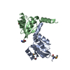
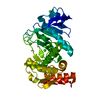
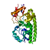
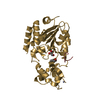
| Title | Aromatic interactions drive the coupled folding and binding of the intrinsically disordered Sesbania mosaic virus VPg protein | ||||||
 Components Components | (Polyprotein Proteolysis) x 2 Proteolysis) x 2 | ||||||
 Keywords Keywords | HYDROLASE / Viral-Protein-genome-linked / complex / protease / PLANT VIRAL PROTEIN | ||||||
| Function / homology |  Function and homology information Function and homology informationhost cell membrane / viral process / serine-type endopeptidase activity / proteolysis / membraneSimilarity search - Function | ||||||
| Biological species |  Sesbania mosaic virus Sesbania mosaic virus | ||||||
| Method | SOLUTION NMR / simulated annealing | ||||||
 Authors Authors | Dixit, K. / Karanth, N.M. / Nair, S. / Kumari, K. / Chakraborti, K.S. / Savithri, H.S. / Sarma, S.P. | ||||||
 Citation Citation | Journal: Biochemistry / Year: 2020 Title: Aromatic Interactions Drive the Coupled Folding and Binding of the Intrinsically Disordered Sesbania mosaic Virus VPg Protein. Authors: Dixit, K. / Karanth, N.M. / Nair, S. / Kumari, K. / Chakrabarti, K.S. / Savithri, H.S. / Sarma, S.P. | ||||||
| History |
|
- Structure visualization
| Structure viewer | Molecule: MolmilJmol/JSmol |
|---|
- Downloads & links
-Download
| PDBx/mmCIF format | 6m78.cif.gz | 195.6 KB | Display | PDBx/mmCIF format |
|---|---|---|---|---|
| PDB format | pdb6m78.ent.gz | 161 KB | Display | PDB format |
| PDBx/mmJSON format | 6m78.json.gz | Tree view | PDBx/mmJSON format | |
| Others |  Other downloads Other downloads |
-Validation report
| Arichive directory | https://data.pdbj.org/pub/pdb/validation_reports/m7/6m78ftp://data.pdbj.org/pub/pdb/validation_reports/m7/6m78 | HTTPS FTP |
|---|
-Related structure data


-Links
 PDBj
PDBj- Assembly
| Deposited unit | 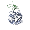
| |||||||||
|---|---|---|---|---|---|---|---|---|---|---|
| 1 |
| |||||||||
| NMR ensembles |
|
-Components
| #1: Protein | Proteolysis Mass: 32245.457 Da / Num. of mol.: 1 Source method: isolated from a genetically manipulated source Source: (gene. exp.) Sesbania mosaic virus / Plasmid: pRSET C / Production host:  Escherichia coli BL21(DE3) (bacteria) / References: UniProt: Q9EB08 Escherichia coli BL21(DE3) (bacteria) / References: UniProt: Q9EB08 |
|---|---|
| #2: Protein | Proteolysis Mass: 9113.224 Da / Num. of mol.: 1 Source method: isolated from a genetically manipulated source Source: (gene. exp.) Sesbania mosaic virus / Plasmid: pET21a / Production host: Escherichia coli BL21(DE3) (bacteria) / Strain (production host): BL21(DE3) / References: UniProt: Q9EB08 |
-Experimental details
-Experiment
| Experiment | Method: SOLUTION NMR | ||||||||||||||||||||||||||||||||||||||||||||||||||||||||||||||||||||||||||||||||||||||||||||||||||||||
|---|---|---|---|---|---|---|---|---|---|---|---|---|---|---|---|---|---|---|---|---|---|---|---|---|---|---|---|---|---|---|---|---|---|---|---|---|---|---|---|---|---|---|---|---|---|---|---|---|---|---|---|---|---|---|---|---|---|---|---|---|---|---|---|---|---|---|---|---|---|---|---|---|---|---|---|---|---|---|---|---|---|---|---|---|---|---|---|---|---|---|---|---|---|---|---|---|---|---|---|---|---|---|---|
| NMR experiment |
|
- Sample preparation
| Details |
|
|---|