 Movie
Movie Controller

[English] 日本語
 Yorodumi
- PDB-3ic5: N-terminal domain of putative saccharopine dehydrogenase from Rue... -
+ Open data

- Basic information
| Entry | Database: PDB / ID: 3ic5 | ||||||
|---|---|---|---|---|---|---|---|
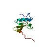
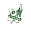
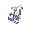
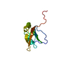
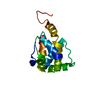
| Title | N-terminal domain of putative saccharopine dehydrogenase from Ruegeria pomeroyi. | ||||||
 Components Components | putative saccharopine dehydrogenase | ||||||
 Keywords Keywords | structural genomics / unknown function / APC63807.2 / N-terminal domain / saccharopine dehydrogenase / PSI-2 / Protein Structure Initiative / Midwest Center for Structural Genomics / MCSG | ||||||
| Function / homology |  Function and homology information Function and homology information | ||||||
| Biological species |  Ruegeria pomeroyi (bacteria) Ruegeria pomeroyi (bacteria) | ||||||
| Method | X-RAY DIFFRACTION / SYNCHROTRON / SAD / Resolution: 2.08 Å | ||||||
 Authors Authors | Osipiuk, J. / Tesar, C. / Freeman, L. / Joachimiak, A. / Midwest Center for Structural Genomics (MCSG) | ||||||
 Citation Citation | Journal: To be Published Title: X-ray crystal structure of N-terminal domain of putative saccharopine dehydrogenase from Ruegeria pomeroyi. Authors: Osipiuk, J. / Tesar, C. / Freeman, L. / Joachimiak, A. | ||||||
| History |
|
- Structure visualization
| Structure viewer | Molecule: MolmilJmol/JSmol |
|---|
- Downloads & links
-Download
| PDBx/mmCIF format | 3ic5.cif.gz | 52.7 KB | Display | PDBx/mmCIF format |
|---|---|---|---|---|
| PDB format | pdb3ic5.ent.gz | 42.2 KB | Display | PDB format |
| PDBx/mmJSON format | 3ic5.json.gz | Tree view | PDBx/mmJSON format | |
| Others |  Other downloads Other downloads |
-Validation report
| Arichive directory | https://data.pdbj.org/pub/pdb/validation_reports/ic/3ic5ftp://data.pdbj.org/pub/pdb/validation_reports/ic/3ic5 | HTTPS FTP |
|---|
-Related structure data
| Similar structure data | |
|---|---|
| Other databases |


-Links
 PDBj
PDBj- Assembly
| Deposited unit | 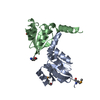
| ||||||||
|---|---|---|---|---|---|---|---|---|---|
| 1 | 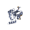
| ||||||||
| 2 | 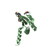
| ||||||||
| Unit cell |
| ||||||||
| Details | putative biological unit is a monomer |
-Components
| #1: Protein | Mass: 12301.582 Da / Num. of mol.: 2 / Fragment: N-terminal domain 1-115 / Mutation: A110V Source method: isolated from a genetically manipulated source Source: (gene. exp.) Ruegeria pomeroyi (bacteria) / Strain: DSS-3 / Gene: SPO0234 / Plasmid: pMCSG19 / Production host: Escherichia coli BL21(DE3) (bacteria) / Strain (production host): BL21(DE3) / References: UniProt: Q5LX24#2: Water | ChemComp-HOH / | Water Mass: 18.015 Da / Num. of mol.: 118 / Source method: isolated from a natural source / Formula: H2O Mass: 18.015 Da / Num. of mol.: 118 / Source method: isolated from a natural source / Formula: H2O |
|---|
-Experimental details
-Experiment
| Experiment | Method: X-RAY DIFFRACTION / Number of used crystals: 1 |
|---|
- Sample preparation
| Crystal | Density Matthews: 2.34 Å3/Da / Density % sol: 47.37 % |
|---|---|
| Crystal grow | Temperature: 277 K / Method: vapor diffusion, sitting drop / pH: 8.5 Details: 0.1 M Tris buffer, 20% ethanol, 5% PEG-400, pH 8.5, VAPOR DIFFUSION, SITTING DROP, temperature 277K |
-Data collection
| Diffraction | Mean temperature: 100 K |
|---|---|
| Diffraction source | Source: SYNCHROTRON / Site: APS  / Beamline: 19-ID / Wavelength: 0.9792 Å / Beamline: 19-ID / Wavelength: 0.9792 Å |
| Detector | Type: ADSC QUANTUM 315 / Detector: CCD / Date: May 30, 2009 |
| Radiation | Monochromator: double crystal monochromator / Protocol: SINGLE WAVELENGTH / Monochromatic (M) / Laue (L): M / Scattering type: x-ray |
| Radiation wavelength | Wavelength: 0.9792 Å / Relative weight: 1 |
| Reflection | Resolution: 2.08→45.4 Å / Num. all: 14419 / Num. obs: 14419 / % possible obs: 100 % / Observed criterion σ(F): 0 / Observed criterion σ(I): 0 / Redundancy: 12.3 % / Biso Wilson estimate: 42.3 Å2 / Rmerge(I) obs: 0.085 / Χ2: 2.457 / Net I/σ(I): 12.7 |
| Reflection shell | Resolution: 2.08→2.12 Å / Redundancy: 7.2 % / Rmerge(I) obs: 0.786 / Mean I/σ(I) obs: 3.22 / Num. unique all: 691 / Χ2: 1.649 / % possible all: 100 |
- Processing
| Software |
| |||||||||||||||||||||||||||||||||||||||||||||||||||||||||||||||||||||||||||||||||||||
|---|---|---|---|---|---|---|---|---|---|---|---|---|---|---|---|---|---|---|---|---|---|---|---|---|---|---|---|---|---|---|---|---|---|---|---|---|---|---|---|---|---|---|---|---|---|---|---|---|---|---|---|---|---|---|---|---|---|---|---|---|---|---|---|---|---|---|---|---|---|---|---|---|---|---|---|---|---|---|---|---|---|---|---|---|---|---|
| Refinement | Method to determine structure: SAD / Resolution: 2.08→45.36 Å / Cor.coef. Fo:Fc: 0.955 / Cor.coef. Fo:Fc free: 0.928 / Occupancy max: 1 / Occupancy min: 0.2 / SU B: 11.678 / SU ML: 0.14 / TLS residual ADP flag: LIKELY RESIDUAL / Cross valid method: THROUGHOUT / σ(F): 0 / σ(I): 0 / ESU R: 0.22 / ESU R Free: 0.194 / Stereochemistry target values: MAXIMUM LIKELIHOOD Details: HYDROGENS HAVE BEEN ADDED IN THE RIDING POSITIONS U VALUES : RESIDUAL ONLY
| |||||||||||||||||||||||||||||||||||||||||||||||||||||||||||||||||||||||||||||||||||||
| Solvent computation | Ion probe radii: 0.8 Å / Shrinkage radii: 0.8 Å / VDW probe radii: 1.2 Å / Solvent model: MASK | |||||||||||||||||||||||||||||||||||||||||||||||||||||||||||||||||||||||||||||||||||||
| Displacement parameters | Biso max: 59.8 Å2 / Biso mean: 28.092 Å2 / Biso min: 15.27 Å2
| |||||||||||||||||||||||||||||||||||||||||||||||||||||||||||||||||||||||||||||||||||||
| Refinement step | Cycle: LAST / Resolution: 2.08→45.36 Å
| |||||||||||||||||||||||||||||||||||||||||||||||||||||||||||||||||||||||||||||||||||||
| Refine LS restraints |
| |||||||||||||||||||||||||||||||||||||||||||||||||||||||||||||||||||||||||||||||||||||
| LS refinement shell | Resolution: 2.08→2.134 Å / Total num. of bins used: 20
| |||||||||||||||||||||||||||||||||||||||||||||||||||||||||||||||||||||||||||||||||||||
| Refinement TLS params. | Method: refined / Refine-ID: X-RAY DIFFRACTION
| |||||||||||||||||||||||||||||||||||||||||||||||||||||||||||||||||||||||||||||||||||||
| Refinement TLS group |
|
