[English] 日本語
Yorodumi
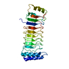
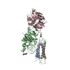
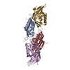
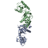
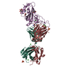
- PDB-6lol: The crystal structure of full length IpaH9.8 -

+
Open data


ID or keywords:

Loading...

-
Basic information

Entry
Database: PDB / ID: 6lol
TitleThe crystal structure of full length IpaH9.8
ComponentsE3 ubiquitin-protein ligase ipaH9.8
KeywordsTRANSFERASE / E3 / IpaH9.8
Function / homology
Function and homology information


modulation of process of another organism / protein K27-linked ubiquitination / host cell cytosol / RING-type E3 ubiquitin transferase / ubiquitin-protein transferase activity / host cell nucleus / extracellular region
Similarity search - Function
Novel E3 ligase domain / C-terminal novel E3 ligase, LRR-interacting / LRR-containing bacterial E3 ligase, N-terminal / Type III secretion system leucine rich repeat protein / Leucine-rich repeat domain superfamily
Similarity search - Domain/homology
E3 ubiquitin-protein ligase ipaH9.8
Similarity search - Component
Biological speciesShigella flexneri 2002017 (bacteria)
MethodX-RAY DIFFRACTION / SYNCHROTRON / MOLECULAR REPLACEMENT / Resolution: 2.75 Å
AuthorsYe, Y. / Huang, H.
Funding support China, 1items
OrganizationGrant numberCountry
National Natural Science Foundation of China (NSFC)21907006 China
CitationJournal: Commun Biol / Year: 2020
Title: Substrate-binding destabilizes the hydrophobic cluster to relieve the autoinhibition of bacterial ubiquitin ligase IpaH9.8.
Authors: Ye, Y. / Xiong, Y. / Huang, H.
History
DepositionJan 6, 2020Deposition site: PDBJ / Processing site: PDBJ
Revision 1.0Dec 23, 2020Provider: repository / Type: Initial release
Revision 1.1Dec 30, 2020Group: Database references / Category: citation
Item: _citation.journal_volume / _citation.page_first ..._citation.journal_volume / _citation.page_first / _citation.page_last / _citation.pdbx_database_id_PubMed / _citation.title
Revision 1.2Nov 29, 2023Group: Data collection / Database references / Refinement description
Category: chem_comp_atom / chem_comp_bond ...chem_comp_atom / chem_comp_bond / database_2 / pdbx_initial_refinement_model
Item: _database_2.pdbx_DOI / _database_2.pdbx_database_accession

-
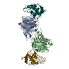
Structure visualization

Structure viewerMolecule:
MolmilJmol/JSmol

Downloads & links

-
Assembly

Deposited unit
A: E3 ubiquitin-protein ligase ipaH9.8


Theoretical massNumber of molelcules
Total (without water)59,9231
Polymers59,9231
Non-polymers00
Water0
1


  • Idetical with deposited unit
  • defined by author
  • Evidence: gel filtration
TypeNameSymmetry operationNumber
identity operation1_555x,y,z1
Buried area0 Å2
ΔGint0 kcal/mol
Surface area25070 Å2
2
A: E3 ubiquitin-protein ligase ipaH9.8

A: E3 ubiquitin-protein ligase ipaH9.8


Theoretical massNumber of molelcules
Total (without water)119,8452
Polymers119,8452
Non-polymers00
Water0
TypeNameSymmetry operationNumber
identity operation1_555x,y,z1
crystal symmetry operation2_555-x,-y,z1
Buried area2180 Å2
ΔGint-2 kcal/mol
Surface area47960 Å2
MethodPISA
Unit cell
Length a, b, c (Å)106.640, 122.250, 149.970
Angle α, β, γ (deg.)90.000, 90.000, 90.000
Int Tables number23
Space group name H-MI222

-
Components

#1: Protein E3 ubiquitin-protein ligase ipaH9.8 / Invasion plasmid antigen ipaH9.8 / RING-type E3 ubiquitin transferase ipaH9.8


Mass: 59922.637 Da / Num. of mol.: 1
Source method: isolated from a genetically manipulated source
Source: (gene. exp.) Shigella flexneri 2002017 (bacteria) / Strain: 2002017 / Gene: ipaH9.8, SFxv_5076 / Production host: Escherichia coli (E. coli)
References: UniProt: D2AJU0, RING-type E3 ubiquitin transferase

-
Experimental details

-
Experiment

ExperimentMethod: X-RAY DIFFRACTION / Number of used crystals: 1

-
Sample preparation

CrystalDensity Matthews: 4.09 Å3/Da / Density % sol: 73.24 %
Crystal growTemperature: 293 K / Method: vapor diffusion, sitting drop / pH: 6.5 / Details: 3.0M NaCl, 0.1M Na Citrate, pH 6.5

-
Data collection

DiffractionMean temperature: 100 K / Serial crystal experiment: N
Diffraction sourceSource: SYNCHROTRON / Site: SSRF / Beamline: BL17U1 / Wavelength: 0.97 Å
DetectorType: DECTRIS EIGER X 16M / Detector: PIXEL / Date: May 17, 2019
RadiationProtocol: SINGLE WAVELENGTH / Monochromatic (M) / Laue (L): M / Scattering type: x-ray
Radiation wavelengthWavelength: 0.97 Å / Relative weight: 1
ReflectionResolution: 2.75→86.91 Å / Num. obs: 25746 / % possible obs: 99.46 % / Redundancy: 6.6 % / Biso Wilson estimate: 81.01 Å2 / CC1/2: 0.994 / CC star: 0.999 / Rmerge(I) obs: 0.08 / Rrim(I) all: 0.0873 / Net I/σ(I): 15.74
Reflection shellResolution: 2.75→2.848 Å / Redundancy: 7 % / Mean I/σ(I) obs: 2.56 / Num. unique obs: 2524 / CC1/2: 0.862 / CC star: 0.962 / % possible all: 99.41

-
Processing

Software
NameVersionClassification
PHENIX1.17.1_3660refinement
Aimlessdata scaling
PDB_EXTRACT3.25data extraction
xia2data reduction
PHENIXphasing
RefinementMethod to determine structure: MOLECULAR REPLACEMENT
Starting model: 5B0T
Resolution: 2.75→86.91 Å / SU ML: 0.39 / Cross valid method: THROUGHOUT / σ(F): 1.34 / Phase error: 34.28 / Stereochemistry target values: ML
RfactorNum. reflection% reflection
Rfree0.2849 1318 5.13 %
Rwork0.2461 24385 -
obs0.248 25703 99.46 %
Solvent computationShrinkage radii: 0.9 Å / VDW probe radii: 1.11 Å / Solvent model: FLAT BULK SOLVENT MODEL
Displacement parametersBiso max: 211.09 Å2 / Biso mean: 102.7644 Å2 / Biso min: 40.08 Å2
Refinement stepCycle: final / Resolution: 2.75→86.91 Å
ProteinNucleic acidLigandSolventTotal
Num. atoms3460 0 0 0 3460
Num. residues----466
LS refinement shell

Refine-ID: X-RAY DIFFRACTION / Rfactor Rfree error: 0

Resolution (Å)Rfactor RfreeNum. reflection RfreeRfactor RworkNum. reflection Rwork% reflection obs (%)
2.75-2.860.37421370.4044251399
2.86-2.990.33941580.31962664100
2.99-3.150.38491430.29512701100
3.15-3.340.28881490.26992683100
3.35-3.60.26241500.2519266999
3.6-3.970.24961230.21572726100
3.97-4.540.23241560.2112705100
4.54-5.720.25641470.23122747100
5.72-86.910.321550.25283699

+
About Yorodumi

-
News

-
Feb 9, 2022. New format data for meta-information of EMDB entries

New format data for meta-information of EMDB entries

  • Version 3 of the EMDB header file is now the official format.
  • The previous official version 1.9 will be removed from the archive.

Related info.:EMDB header

External links:wwPDB to switch to version 3 of the EMDB data model

-
Aug 12, 2020. Covid-19 info

Covid-19 info

URL: https://pdbj.org/emnavi/covid19.php

New page: Covid-19 featured information page in EM Navigator.

Related info.:Covid-19 info / Mar 5, 2020. Novel coronavirus structure data

+
Mar 5, 2020. Novel coronavirus structure data

Novel coronavirus structure data

Related info.:Yorodumi Speices / Aug 12, 2020. Covid-19 info

External links:COVID-19 featured content - PDBj / Molecule of the Month (242):Coronavirus Proteases

+
Jan 31, 2019. EMDB accession codes are about to change! (news from PDBe EMDB page)

EMDB accession codes are about to change! (news from PDBe EMDB page)

  • The allocation of 4 digits for EMDB accession codes will soon come to an end. Whilst these codes will remain in use, new EMDB accession codes will include an additional digit and will expand incrementally as the available range of codes is exhausted. The current 4-digit format prefixed with “EMD-” (i.e. EMD-XXXX) will advance to a 5-digit format (i.e. EMD-XXXXX), and so on. It is currently estimated that the 4-digit codes will be depleted around Spring 2019, at which point the 5-digit format will come into force.
  • The EM Navigator/Yorodumi systems omit the EMD- prefix.

Related info.:Q: What is EMD? / ID/Accession-code notation in Yorodumi/EM Navigator

External links:EMDB Accession Codes are Changing Soon! / Contact to PDBj

+
Jul 12, 2017. Major update of PDB

Major update of PDB

  • wwPDB released updated PDB data conforming to the new PDBx/mmCIF dictionary.
  • This is a major update changing the version number from 4 to 5, and with Remediation, in which all the entries are updated.
  • In this update, many items about electron microscopy experimental information are reorganized (e.g. em_software).
  • Now, EM Navigator and Yorodumi are based on the updated data.

External links:wwPDB Remediation / Enriched Model Files Conforming to OneDep Data Standards Now Available in the PDB FTP Archive

-
Yorodumi

Thousand views of thousand structures

  • Yorodumi is a browser for structure data from EMDB, PDB, SASBDB, etc.
  • This page is also the successor to EM Navigator detail page, and also detail information page/front-end page for Omokage search.
  • The word "yorodu" (or yorozu) is an old Japanese word meaning "ten thousand". "mi" (miru) is to see.

Related info.:EMDB / PDB / SASBDB / Comparison of 3 databanks / Yorodumi Search / Aug 31, 2016. New EM Navigator & Yorodumi / Yorodumi Papers / Jmol/JSmol / Function and homology information / Changes in new EM Navigator and Yorodumi

Read more