 Movie
Movie Controller


+ Open data

- Basic information
| Entry | Database: PDB / ID: 3cvr | ||||||
|---|---|---|---|---|---|---|---|
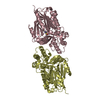
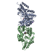
| Title | Crystal structure of the full length IpaH3 | ||||||
 Components Components | Invasion plasmid antigen | ||||||
 Keywords Keywords |  LIGASE / Leucine rich repeat and alpha fold LIGASE / Leucine rich repeat and alpha fold | ||||||
| Function / homology |  Function and homology information Function and homology information: / RING-type E3 ubiquitin transferase / ubiquitin-protein transferase activity / host cell cytoplasm / extracellular region Similarity search - Function | ||||||
| Biological species |  Shigella flexneri 2a (bacteria) Shigella flexneri 2a (bacteria) | ||||||
| Method | X-RAY DIFFRACTION / SYNCHROTRON / SAD / Resolution: 2.8 Å | ||||||
 Authors Authors | Zhu, Y. / Shao, F. | ||||||
 Citation Citation | Journal: Nat.Struct.Mol.Biol. / Year: 2008 Title: Structure of a Shigella effector reveals a new class of ubiquitin ligases Authors: Zhu, Y. / Li, H. / Hu, L. / Wang, J. / Zhou, Y. / Pang, Z. / Liu, L. / Shao, F. | ||||||
| History |
|
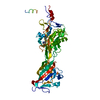
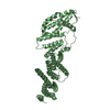
- Structure visualization
| Structure viewer | Molecule: MolmilJmol/JSmol |
|---|
- Downloads & links
-Download
| PDBx/mmCIF format | 3cvr.cif.gz | 102.2 KB | Display | PDBx/mmCIF format |
|---|---|---|---|---|
| PDB format | pdb3cvr.ent.gz | 81.7 KB | Display | PDB format |
| PDBx/mmJSON format | 3cvr.json.gz | Tree view | PDBx/mmJSON format | |
| Others |  Other downloads Other downloads |
-Validation report
| Arichive directory | https://data.pdbj.org/pub/pdb/validation_reports/cv/3cvrftp://data.pdbj.org/pub/pdb/validation_reports/cv/3cvr | HTTPS FTP |
|---|
-Related structure data
| Similar structure data |
|---|


-Links
 PDBj
PDBj


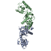
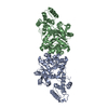
- Assembly
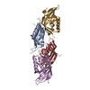
| Deposited unit | 
| ||||||||
|---|---|---|---|---|---|---|---|---|---|
| 1 |
| ||||||||
| 2 | 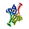
| ||||||||
| Unit cell |
|
-Components
| #1: Protein | Mass: 65508.297 Da / Num. of mol.: 1 Source method: isolated from a genetically manipulated source Source: (gene. exp.) Shigella flexneri 2a (bacteria) / Strain: 301 / Gene: ipaH_3 / Plasmid: pGEX-6p-2 / Production host: Escherichia coli (E. coli) / Strain (production host): Bl21(DE3)References: UniProt: Q83RJ4, Ligases; Forming carbon-nitrogen bonds; Acid-amino-acid ligases (peptide synthases) |
|---|---|
| #2: Water | ChemComp-HOH / Water Mass: 18.015 Da / Num. of mol.: 39 / Source method: isolated from a natural source / Formula: H2O Mass: 18.015 Da / Num. of mol.: 39 / Source method: isolated from a natural source / Formula: H2O |
| Sequence details | THERE ARE CONFLICTS BETWEEN THE CLONED SEQUENCE IN THIS ENTRY AND THE DATABASE REFERENCE SEQUENCE. ...THERE ARE CONFLICTS BETWEEN THE CLONED SEQUENCE IN THIS ENTRY AND THE DATABASE REFERENCE SEQUENCE. THE DEPOSITOR SUGGESTS THE SEQUENCE CONFLICTS ARE NOT THE MUTATIONS BUT MAYBE THE REAL RESIDUES IN THE STAIN. |
-Experimental details
-Experiment
| Experiment | Method: X-RAY DIFFRACTION / Number of used crystals: 2 |
|---|
- Sample preparation
| Crystal | Density Matthews: 3.9 Å3/Da / Density % sol: 68.43 % |
|---|---|
| Crystal grow | Temperature: 293 K / Method: vapor diffusion, hanging drop / pH: 5.5 Details: 2M NaCl, 0.1M Bis-Tris pH5.5, 2.5% Polyacrylic acid 5100 sodium salt, 0.1 M Sodium Nitrate, VAPOR DIFFUSION, HANGING DROP, temperature 293K |
-Data collection
| Diffraction |
| |||||||||||||||
|---|---|---|---|---|---|---|---|---|---|---|---|---|---|---|---|---|
| Diffraction source |
| |||||||||||||||
| Detector |
| |||||||||||||||
| Radiation |
| |||||||||||||||
| Radiation wavelength |
| |||||||||||||||
| Reflection | Resolution: 2.8→51.37 Å / Num. obs: 26107 / % possible obs: 98.7 % / Observed criterion σ(F): 1 / Observed criterion σ(I): 1 / Redundancy: 11 % / Rmerge(I) obs: 0.071 / Rsym value: 0.074 / Net I/σ(I): 25.3 | |||||||||||||||
| Reflection shell | Resolution: 2.8→2.95 Å / Redundancy: 11.1 % / Rmerge(I) obs: 0.362 / Mean I/σ(I) obs: 5.5 / Rsym value: 0.38 / % possible all: 99.6 |


-Phasing
| Phasing | Method: SAD |
|---|
- Processing
| Software |
| ||||||||||||||||||||||||||||||||||||||||||||||||||||||||||||||||||||||||||||||||||||||||||||||||||||||||||||||||||||||||||||||||||||||||||||||||||||||||||||||||||||||||||
|---|---|---|---|---|---|---|---|---|---|---|---|---|---|---|---|---|---|---|---|---|---|---|---|---|---|---|---|---|---|---|---|---|---|---|---|---|---|---|---|---|---|---|---|---|---|---|---|---|---|---|---|---|---|---|---|---|---|---|---|---|---|---|---|---|---|---|---|---|---|---|---|---|---|---|---|---|---|---|---|---|---|---|---|---|---|---|---|---|---|---|---|---|---|---|---|---|---|---|---|---|---|---|---|---|---|---|---|---|---|---|---|---|---|---|---|---|---|---|---|---|---|---|---|---|---|---|---|---|---|---|---|---|---|---|---|---|---|---|---|---|---|---|---|---|---|---|---|---|---|---|---|---|---|---|---|---|---|---|---|---|---|---|---|---|---|---|---|---|---|---|---|
| Refinement | Method to determine structure: SAD / Resolution: 2.8→48.76 Å / Cor.coef. Fo:Fc: 0.909 / Cor.coef. Fo:Fc free: 0.888 / SU B: 25.245 / SU ML: 0.243 / TLS residual ADP flag: LIKELY RESIDUAL / Cross valid method: THROUGHOUT / ESU R: 0.446 / ESU R Free: 0.308 / Stereochemistry target values: MAXIMUM LIKELIHOOD / Details: HYDROGENS HAVE BEEN ADDED IN THE RIDING POSITIONS
| ||||||||||||||||||||||||||||||||||||||||||||||||||||||||||||||||||||||||||||||||||||||||||||||||||||||||||||||||||||||||||||||||||||||||||||||||||||||||||||||||||||||||||
| Solvent computation | Ion probe radii: 0.8 Å / Shrinkage radii: 0.8 Å / VDW probe radii: 1.2 Å / Solvent model: MASK | ||||||||||||||||||||||||||||||||||||||||||||||||||||||||||||||||||||||||||||||||||||||||||||||||||||||||||||||||||||||||||||||||||||||||||||||||||||||||||||||||||||||||||
| Displacement parameters | Biso mean: 68.787 Å2
| ||||||||||||||||||||||||||||||||||||||||||||||||||||||||||||||||||||||||||||||||||||||||||||||||||||||||||||||||||||||||||||||||||||||||||||||||||||||||||||||||||||||||||
| Refinement step | Cycle: LAST / Resolution: 2.8→48.76 Å
| ||||||||||||||||||||||||||||||||||||||||||||||||||||||||||||||||||||||||||||||||||||||||||||||||||||||||||||||||||||||||||||||||||||||||||||||||||||||||||||||||||||||||||
| Refine LS restraints |
| ||||||||||||||||||||||||||||||||||||||||||||||||||||||||||||||||||||||||||||||||||||||||||||||||||||||||||||||||||||||||||||||||||||||||||||||||||||||||||||||||||||||||||
| LS refinement shell | Resolution: 2.8→2.873 Å / Total num. of bins used: 20
| ||||||||||||||||||||||||||||||||||||||||||||||||||||||||||||||||||||||||||||||||||||||||||||||||||||||||||||||||||||||||||||||||||||||||||||||||||||||||||||||||||||||||||
| Refinement TLS params. | Method: refined / Refine-ID: X-RAY DIFFRACTION
| ||||||||||||||||||||||||||||||||||||||||||||||||||||||||||||||||||||||||||||||||||||||||||||||||||||||||||||||||||||||||||||||||||||||||||||||||||||||||||||||||||||||||||
| Refinement TLS group |
| ||||||||||||||||||||||||||||||||||||||||||||||||||||||||||||||||||||||||||||||||||||||||||||||||||||||||||||||||||||||||||||||||||||||||||||||||||||||||||||||||||||||||||
| Xplor file |
|
