 Movie
Movie Controller


+ Open data

- Basic information
| Entry | Database: PDB / ID: 5b0t | ||||||
|---|---|---|---|---|---|---|---|
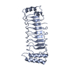
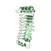
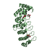
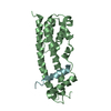
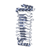
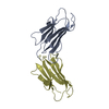
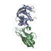
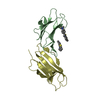
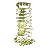
| Title | Structure of Shigella effector LRR domain | ||||||
 Components Components | E3 ubiquitin-protein ligase ipaH9.8 | ||||||
 Keywords Keywords |  LIGASE / effector / ubiquitin ligase / LRR domain LIGASE / effector / ubiquitin ligase / LRR domain | ||||||
| Function / homology |  Function and homology information Function and homology informationsymbiont-mediated suppression of host inflammatory response / symbiont-mediated suppression of host NF-kappaB cascade / symbiont-mediated suppression of host innate immune response / symbiont-mediated suppression of host defenses / protein K27-linked ubiquitination / host cell cytosol / protein autoubiquitination / protein K48-linked ubiquitination / RING-type E3 ubiquitin transferase / ubiquitin-protein transferase activity ...symbiont-mediated suppression of host inflammatory response / symbiont-mediated suppression of host NF-kappaB cascade / symbiont-mediated suppression of host innate immune response / symbiont-mediated suppression of host defenses / protein K27-linked ubiquitination / host cell cytosol / protein autoubiquitination / protein K48-linked ubiquitination / RING-type E3 ubiquitin transferase / ubiquitin-protein transferase activity / ubiquitin protein ligase activity / ubiquitin-dependent protein catabolic process / proteasome-mediated ubiquitin-dependent protein catabolic process / host cell nucleus / extracellular region / identical protein bindingSimilarity search - Function | ||||||
| Biological species |  Shigella flexneri (bacteria) Shigella flexneri (bacteria) | ||||||
| Method | X-RAY DIFFRACTION / SYNCHROTRON / MOLECULAR REPLACEMENT / molecular replacement / Resolution: 2 Å | ||||||
 Authors Authors | Takagi, K. / Sasakawa, C. / Kim, M. / Mizushima, T. | ||||||
 Citation Citation | Journal: Acta Crystallogr.,Sect.F / Year: 2016 Title: Crystal structure of the substrate-recognition domain of the Shigella E3 ligase IpaH9.8 Authors: Takagi, K. / Kim, M. / Sasakawa, C. / Mizushima, T. | ||||||
| History |
|
- Structure visualization
| Structure viewer | Molecule: MolmilJmol/JSmol |
|---|
- Downloads & links
-Download
| PDBx/mmCIF format | 5b0t.cif.gz | 57.7 KB | Display | PDBx/mmCIF format |
|---|---|---|---|---|
| PDB format | pdb5b0t.ent.gz | 41.4 KB | Display | PDB format |
| PDBx/mmJSON format | 5b0t.json.gz | Tree view | PDBx/mmJSON format | |
| Others |  Other downloads Other downloads |
-Validation report
| Arichive directory | https://data.pdbj.org/pub/pdb/validation_reports/b0/5b0tftp://data.pdbj.org/pub/pdb/validation_reports/b0/5b0t | HTTPS FTP |
|---|
-Related structure data

-Links
 PDBj
PDBj- Assembly
| Deposited unit | 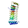
| ||||||||
|---|---|---|---|---|---|---|---|---|---|
| 1 |
| ||||||||
| Unit cell |
|
-Components
| #1: Protein | Mass: 25377.514 Da / Num. of mol.: 1 / Fragment: UNP residues 21-244 Source method: isolated from a genetically manipulated source Source: (gene. exp.) Shigella flexneri (bacteria) / Gene: ipaH9.8, CP0226, pWR501_0234, SFLP090 / Plasmid: pCold1 / Production host: Escherichia coli BL21(DE3) (bacteria) / Strain (production host): BL21(DE3)References: UniProt: Q8VSC3, Ligases; Forming carbon-nitrogen bonds; Acid-amino-acid ligases (peptide synthases) |
|---|---|
| #2: Water | ChemComp-HOH / Water Mass: 18.015 Da / Num. of mol.: 52 / Source method: isolated from a natural source / Formula: H2O Mass: 18.015 Da / Num. of mol.: 52 / Source method: isolated from a natural source / Formula: H2O |
-Experimental details
-Experiment
| Experiment | Method: X-RAY DIFFRACTION / Number of used crystals: 1 |
|---|
- Sample preparation
| Crystal | Density Matthews: 2.18 Å3/Da / Density % sol: 43.45 % |
|---|---|
| Crystal grow | Temperature: 293 K / Method: vapor diffusion, sitting drop / pH: 5.5 Details: 0.1M BIS-TRIS pH5.5, 0.2M Ammonium Sulfate, 25% (w/v) PEG3350 |
-Data collection
| Diffraction | Mean temperature: 100 K |
|---|---|
| Diffraction source | Source: SYNCHROTRON / Site: SPring-8  / Beamline: BL44XU / Wavelength: 0.9 Å / Beamline: BL44XU / Wavelength: 0.9 Å |
| Detector | Type: RAYONIX MX300HE / Detector: CCD / Date: Jun 18, 2014 |
| Radiation | Protocol: SINGLE WAVELENGTH / Monochromatic (M) / Laue (L): M / Scattering type: x-ray |
| Radiation wavelength | Wavelength: 0.9 Å / Relative weight: 1 |
| Reflection | Resolution: 2→50 Å / Num. obs: 15296 / % possible obs: 99.8 % / Redundancy: 6.7 % / Rmerge(I) obs: 0.054 / Net I/σ(I): 44.3 |
| Reflection shell | Resolution: 2→2.03 Å / Redundancy: 6.9 % / % possible all: 100 |
-Phasing
| Phasing | Method: molecular replacement |
|---|
- Processing
| Software |
| |||||||||||||||||||||||||||||||||||||||||||||
|---|---|---|---|---|---|---|---|---|---|---|---|---|---|---|---|---|---|---|---|---|---|---|---|---|---|---|---|---|---|---|---|---|---|---|---|---|---|---|---|---|---|---|---|---|---|---|
| Refinement | Method to determine structure: MOLECULAR REPLACEMENT / Resolution: 2→34.14 Å / Cor.coef. Fo:Fc: 0.948 / Cor.coef. Fo:Fc free: 0.918 / SU B: 5.664 / SU ML: 0.154 / Cross valid method: THROUGHOUT / σ(F): 0 / ESU R: 0.218 / ESU R Free: 0.192 / Stereochemistry target values: MAXIMUM LIKELIHOOD Details: HYDROGENS HAVE BEEN USED IF PRESENT IN THE INPUT U VALUES : REFINED INDIVIDUALLY
| |||||||||||||||||||||||||||||||||||||||||||||
| Solvent computation | Ion probe radii: 0.8 Å / Shrinkage radii: 0.8 Å / VDW probe radii: 1.2 Å / Solvent model: MASK | |||||||||||||||||||||||||||||||||||||||||||||
| Displacement parameters | Biso max: 112.83 Å2 / Biso mean: 35.3 Å2 / Biso min: 11.77 Å2
| |||||||||||||||||||||||||||||||||||||||||||||
| Refinement step | Cycle: final / Resolution: 2→34.14 Å
| |||||||||||||||||||||||||||||||||||||||||||||
| Refine LS restraints |
| |||||||||||||||||||||||||||||||||||||||||||||
| LS refinement shell | Resolution: 2.001→2.053 Å / Total num. of bins used: 20
|
