 Movie
Movie Controller


+ Open data

- Basic information
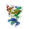
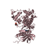
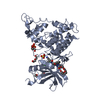
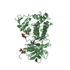
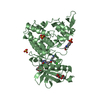
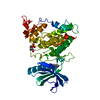
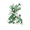
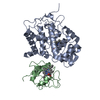
| Entry | Database: PDB / ID: 6ln1 | ||||||
|---|---|---|---|---|---|---|---|
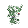
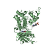
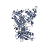
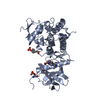
| Title | A natural inhibitor of DYRK1A for treatment of diabetes mellitus | ||||||
 Components Components | Dual specificity tyrosine-phosphorylation-regulated kinase 1A DYRK1A DYRK1A | ||||||
 Keywords Keywords | TRANSFERASE/TRANSFERASE INHIBITOR / anti-diabetic activity / Desmethylbellidifolin / DYRK1A / inhibitor / TRANSFERASE / TRANSFERASE-TRANSFERASE INHIBITOR complex | ||||||
| Function / homology |  Function and homology information Function and homology informationhistone H3T45 kinase activity / positive regulation of protein deacetylation / peptidyl-serine autophosphorylation / negative regulation of DNA methylation-dependent heterochromatin formation / dual-specificity kinase / [RNA-polymerase]-subunit kinase / negative regulation of microtubule polymerization / tau-protein kinase activity / negative regulation of DNA damage response, signal transduction by p53 class mediator / negative regulation of mRNA splicing, via spliceosome ...histone H3T45 kinase activity / positive regulation of protein deacetylation / peptidyl-serine autophosphorylation / negative regulation of DNA methylation-dependent heterochromatin formation / dual-specificity kinase / [RNA-polymerase]-subunit kinase / negative regulation of microtubule polymerization / tau-protein kinase activity / negative regulation of DNA damage response, signal transduction by p53 class mediator / negative regulation of mRNA splicing, via spliceosome / amyloid-beta formation / G0 and Early G1 / peptidyl-tyrosine autophosphorylation / cytoskeletal protein binding / protein serine/threonine/tyrosine kinase activity / tubulin binding / RNA polymerase II CTD heptapeptide repeat kinase activity / positive regulation of RNA splicing / peptidyl-threonine phosphorylation / non-membrane spanning protein tyrosine kinase activity / tau protein binding / circadian rhythm / peptidyl-tyrosine phosphorylation / : / nervous system development / actin binding / peptidyl-serine phosphorylation / protein tyrosine kinase activity / protein autophosphorylation / transcription coactivator activity / cytoskeleton / protein kinase activity / nuclear speck / ribonucleoprotein complex / axon / protein phosphorylation / protein serine kinase activity / protein serine/threonine kinase activity / dendrite / positive regulation of DNA-templated transcription / nucleoplasm / ATP binding / identical protein binding / nucleus / cytoplasmSimilarity search - Function | ||||||
| Biological species |  Homo sapiens (human) Homo sapiens (human) | ||||||
| Method | X-RAY DIFFRACTION / SYNCHROTRON / MOLECULAR REPLACEMENT / molecular replacement / Resolution: 2.699 Å | ||||||
 Authors Authors | Li, H. / Chen, L.X. / Zheng, M.Z. / Zhang, Q.Z. / Zhang, C.L. / Wu, C.R. / Yang, K.Y. / Song, Z.R. / Wang, Q.Q. / Li, C. ...Li, H. / Chen, L.X. / Zheng, M.Z. / Zhang, Q.Z. / Zhang, C.L. / Wu, C.R. / Yang, K.Y. / Song, Z.R. / Wang, Q.Q. / Li, C. / Zhou, Y.R. / Chen, J.C. | ||||||
| Funding support |  China, 1items China, 1items
| ||||||
 Citation Citation | Journal: Clin Transl Med / Year: 2021 Title: A natural DYRK1A inhibitor as a potential stimulator for beta-cell proliferation in diabetes. Authors: Zheng, M. / Zhang, Q. / Zhang, C. / Wu, C. / Yang, K. / Song, Z. / Wang, Q. / Li, C. / Zhou, Y. / Chen, J. / Li, H. / Chen, L. | ||||||
| History |
|
- Structure visualization
| Structure viewer | Molecule: MolmilJmol/JSmol |
|---|
- Downloads & links
-Download
| PDBx/mmCIF format | 6ln1.cif.gz | 152.7 KB | Display | PDBx/mmCIF format |
|---|---|---|---|---|
| PDB format | pdb6ln1.ent.gz | 120 KB | Display | PDB format |
| PDBx/mmJSON format | 6ln1.json.gz | Tree view | PDBx/mmJSON format | |
| Others |  Other downloads Other downloads |
-Validation report
| Arichive directory | https://data.pdbj.org/pub/pdb/validation_reports/ln/6ln1ftp://data.pdbj.org/pub/pdb/validation_reports/ln/6ln1 | HTTPS FTP |
|---|
-Related structure data
| Related structure data | 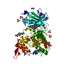 4ylkS S: Starting model for refinement |
|---|---|
| Similar structure data |


-Links
 PDBj
PDBj
- Assembly
| Deposited unit | 
| ||||||||
|---|---|---|---|---|---|---|---|---|---|
| 1 | 
| ||||||||
| 2 | 
| ||||||||
| Unit cell |
|
-Components
| #1: Protein | DYRK1A / Dual specificity YAK1-related kinase / HP86 / Protein kinase minibrain homolog / hMNB Mass: 40478.727 Da / Num. of mol.: 2 Source method: isolated from a genetically manipulated source Source: (gene. exp.) Homo sapiens (human) / Gene: DYRK1A, DYRK, MNB, MNBH / Production host:  Escherichia coli (E. coli) / References: UniProt: Q13627, dual-specificity kinase Escherichia coli (E. coli) / References: UniProt: Q13627, dual-specificity kinase#2: Chemical |   Mass: 260.199 Da / Num. of mol.: 2 / Source method: obtained synthetically / Formula: C13H8O6 / Feature type: SUBJECT OF INVESTIGATION Mass: 260.199 Da / Num. of mol.: 2 / Source method: obtained synthetically / Formula: C13H8O6 / Feature type: SUBJECT OF INVESTIGATION#3: Water | ChemComp-HOH / | Water Mass: 18.015 Da / Num. of mol.: 44 / Source method: isolated from a natural source / Formula: H2O Mass: 18.015 Da / Num. of mol.: 44 / Source method: isolated from a natural source / Formula: H2OHas ligand of interest | Y | |
|---|
-Experimental details
-Experiment
| Experiment | Method: X-RAY DIFFRACTION / Number of used crystals: 1 |
|---|
- Sample preparation
| Crystal | Density Matthews: 3.02 Å3/Da / Density % sol: 59.21 % |
|---|---|
| Crystal grow | Temperature: 277 K / Method: vapor diffusion, sitting drop / Details: 0.1M Bis-Tris, 17% PEG 1500, 30% ethylene glycol |
-Data collection
| Diffraction | Mean temperature: 100 K / Serial crystal experiment: N |
|---|---|
| Diffraction source | Source: SYNCHROTRON / Site: SSRF / Beamline: BL17U1 / Wavelength: 1 Å |
| Detector | Type: DECTRIS PILATUS3 R CdTe 300K / Detector: PIXEL / Date: Apr 6, 2019 |
| Radiation | Protocol: SINGLE WAVELENGTH / Monochromatic (M) / Laue (L): M / Scattering type: x-ray |
| Radiation wavelength | Wavelength: 1 Å / Relative weight: 1 |
| Reflection | Resolution: 2.699→50 Å / Num. obs: 25854 / % possible obs: 100 % / Redundancy: 10.5 % / Biso Wilson estimate: 61.94 Å2 / CC1/2: 0.998 / Rmerge(I) obs: 0.095 / Rpim(I) all: 0.031 / Rrim(I) all: 0.1 / Rsym value: 0.068 / Net I/σ(I): 22.3 |
| Reflection shell | Resolution: 2.7→2.75 Å / Rmerge(I) obs: 1.196 / Mean I/σ(I) obs: 1.52 / Num. unique obs: 1304 / CC1/2: 0.731 / Rpim(I) all: 0.384 / Rrim(I) all: 1.256 / Rsym value: 1.365 / % possible all: 100 |
-Phasing
| Phasing | Method: molecular replacement |
|---|
- Processing
| Software |
| |||||||||||||||||||||||||||||||||||||||||||||||||||||||||||||||||||||||||||
|---|---|---|---|---|---|---|---|---|---|---|---|---|---|---|---|---|---|---|---|---|---|---|---|---|---|---|---|---|---|---|---|---|---|---|---|---|---|---|---|---|---|---|---|---|---|---|---|---|---|---|---|---|---|---|---|---|---|---|---|---|---|---|---|---|---|---|---|---|---|---|---|---|---|---|---|---|
| Refinement | Method to determine structure: MOLECULAR REPLACEMENT Starting model: 4YLK Resolution: 2.699→48.974 Å / SU ML: 0.43 / Cross valid method: THROUGHOUT / σ(F): 1.38 / Phase error: 28.03 / Stereochemistry target values: ML
| |||||||||||||||||||||||||||||||||||||||||||||||||||||||||||||||||||||||||||
| Solvent computation | Shrinkage radii: 0.9 Å / VDW probe radii: 1.11 Å / Solvent model: FLAT BULK SOLVENT MODEL | |||||||||||||||||||||||||||||||||||||||||||||||||||||||||||||||||||||||||||
| Displacement parameters | Biso max: 123.42 Å2 / Biso mean: 65.7556 Å2 / Biso min: 25.89 Å2 | |||||||||||||||||||||||||||||||||||||||||||||||||||||||||||||||||||||||||||
| Refinement step | Cycle: final / Resolution: 2.699→48.974 Å
| |||||||||||||||||||||||||||||||||||||||||||||||||||||||||||||||||||||||||||
| LS refinement shell | Refine-ID: X-RAY DIFFRACTION / Rfactor Rfree error: 0 / % reflection obs: 100 %
|
