| Entry | Database: PDB / ID: 6a1g
|
|---|
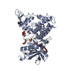
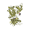
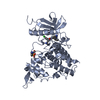
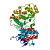
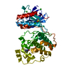
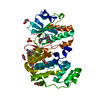
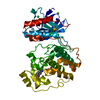
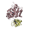
| Title | Crystal structure of human DYRK1A in complex with compound 32 |
|---|
 Components Components | Dual specificity tyrosine-phosphorylation-regulated kinase 1A DYRK1A DYRK1A |
|---|
 Keywords Keywords | TRANSFERASE / DYRK1a |
|---|
| Function / homology |  Function and homology information Function and homology information
histone H3T45 kinase activity / positive regulation of protein deacetylation / peptidyl-serine autophosphorylation / negative regulation of DNA methylation-dependent heterochromatin formation / dual-specificity kinase / [RNA-polymerase]-subunit kinase / negative regulation of microtubule polymerization / tau-protein kinase activity / negative regulation of DNA damage response, signal transduction by p53 class mediator / negative regulation of mRNA splicing, via spliceosome ...histone H3T45 kinase activity / positive regulation of protein deacetylation / peptidyl-serine autophosphorylation / negative regulation of DNA methylation-dependent heterochromatin formation / dual-specificity kinase / [RNA-polymerase]-subunit kinase / negative regulation of microtubule polymerization / tau-protein kinase activity / negative regulation of DNA damage response, signal transduction by p53 class mediator / negative regulation of mRNA splicing, via spliceosome / amyloid-beta formation / G0 and Early G1 / peptidyl-tyrosine autophosphorylation / cytoskeletal protein binding / protein serine/threonine/tyrosine kinase activity / tubulin binding / RNA polymerase II CTD heptapeptide repeat kinase activity / positive regulation of RNA splicing / peptidyl-threonine phosphorylation / non-membrane spanning protein tyrosine kinase activity / tau protein binding / circadian rhythm / peptidyl-tyrosine phosphorylation / : / nervous system development / actin binding / peptidyl-serine phosphorylation / protein tyrosine kinase activity / protein autophosphorylation / transcription coactivator activity / cytoskeleton / protein kinase activity / nuclear speck / ribonucleoprotein complex / axon / protein phosphorylation / protein serine kinase activity / protein serine/threonine kinase activity / dendrite / positive regulation of DNA-templated transcription / nucleoplasm / ATP binding / identical protein binding / nucleus / cytoplasmSimilarity search - Function Dual specificity tyrosine-phosphorylation-regulated kinase 1A/1B, catalytic domain / Transferase(Phosphotransferase) domain 1 / Transferase(Phosphotransferase); domain 1 / Serine/threonine-protein kinase, active site / Serine/Threonine protein kinases active-site signature. / Protein kinase domain / Serine/Threonine protein kinases, catalytic domain / Protein kinase, ATP binding site / Protein kinases ATP-binding region signature. / Protein kinase domain profile. ...Dual specificity tyrosine-phosphorylation-regulated kinase 1A/1B, catalytic domain / Transferase(Phosphotransferase) domain 1 / Transferase(Phosphotransferase); domain 1 / Serine/threonine-protein kinase, active site / Serine/Threonine protein kinases active-site signature. / Protein kinase domain / Serine/Threonine protein kinases, catalytic domain / Protein kinase, ATP binding site / Protein kinases ATP-binding region signature. / Protein kinase domain profile. / Protein kinase domain / Protein kinase-like domain superfamily / Orthogonal Bundle / Mainly AlphaSimilarity search - Domain/homology |
|---|
| Biological species |  Homo sapiens (human) Homo sapiens (human) |
|---|
| Method | X-RAY DIFFRACTION / SYNCHROTRON / MOLECULAR REPLACEMENT / Resolution: 2.15 Å |
|---|
 Authors Authors | Baba, D. / Hanzawa, H. |
|---|
 Citation Citation | Journal: Bioorg. Med. Chem. Lett. / Year: 2018
Title: Discovery of DS42450411 as a potent orally active hepcidin production inhibitor: Design and optimization of novel 4-aminopyrimidine derivatives.
Authors: Fukuda, T. / Ishiyama, T. / Katagiri, T. / Ueda, K. / Muramatsu, S. / Hashimoto, M. / Aki, A. / Baba, D. / Watanabe, K. / Tanaka, N. |
|---|
| History | | Deposition | Jun 7, 2018 | Deposition site: PDBJ / Processing site: PDBJ |
|---|
| Revision 1.0 | Oct 3, 2018 | Provider: repository / Type: Initial release |
|---|
| Revision 1.1 | Oct 24, 2018 | Group: Data collection / Database references / Category: citation
Item: _citation.journal_volume / _citation.page_first / _citation.page_last |
|---|
| Revision 1.2 | Nov 22, 2023 | Group: Data collection / Database references / Refinement description
Category: chem_comp_atom / chem_comp_bond ...chem_comp_atom / chem_comp_bond / database_2 / pdbx_initial_refinement_model / struct_ncs_dom_lim
Item: _database_2.pdbx_DOI / _database_2.pdbx_database_accession ..._database_2.pdbx_DOI / _database_2.pdbx_database_accession / _struct_ncs_dom_lim.beg_auth_comp_id / _struct_ncs_dom_lim.beg_label_asym_id / _struct_ncs_dom_lim.beg_label_comp_id / _struct_ncs_dom_lim.beg_label_seq_id / _struct_ncs_dom_lim.end_auth_comp_id / _struct_ncs_dom_lim.end_label_asym_id / _struct_ncs_dom_lim.end_label_comp_id / _struct_ncs_dom_lim.end_label_seq_id |
|---|
|
|---|
 Movie
Movie Controller


 Open data
 Basic information
 Structure visualization
 Downloads & links
 Other downloads
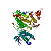


 PDBj
PDBj
 Assembly
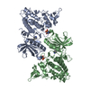


 Mass: 334.458 Da / Num. of mol.: 2 / Source method: obtained synthetically / Formula: C21H26N4 / Feature type: SUBJECT OF INVESTIGATION
 Mass: 18.015 Da / Num. of mol.: 151 / Source method: isolated from a natural source / Formula: H2O
 Sample preparation
 / Beamline: BL-17A / Wavelength: 1 Å
 Processing