 Movie
Movie Controller

[English] 日本語
 Yorodumi
- PDB-6lh1: Crystal structure of a cysteine-pair mutant (Y113C-P190C) of a ba... -
+ Open data

- Basic information
| Entry | Database: PDB / ID: 6lh1 | ||||||
|---|---|---|---|---|---|---|---|
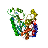
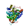
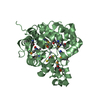
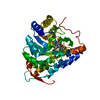
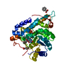
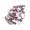
| Title | Crystal structure of a cysteine-pair mutant (Y113C-P190C) of a bacterial bile acid transporter trapped in an outward-facing conformation | ||||||
 Components Components | Transporter, sodium/bile acid symporter family Transport protein Transport protein | ||||||
 Keywords Keywords | TRANSPORT PROTEIN / Bile acid transporter / ASBT / NTCP / SLC10 | ||||||
| Function / homology | Bile acid:sodium symporter/arsenical resistance protein Acr3 / Bile acid:sodium symporter / Sodium Bile acid symporter family / Sodium/solute symporter superfamily / membrane / 2,3-dihydroxypropyl (9Z)-octadec-9-enoate / CITRIC ACID / Transporter, sodium/bile acid symporter family Function and homology information Function and homology information | ||||||
| Biological species |  Yersinia frederiksenii (bacteria) Yersinia frederiksenii (bacteria) | ||||||
| Method | X-RAY DIFFRACTION / SYNCHROTRON / MOLECULAR REPLACEMENT / molecular replacement / Resolution: 2.861 Å | ||||||
 Authors Authors | Wang, X. / Lyu, Y. / Ji, Y. / Sun, Z. / Zhou, X. | ||||||
| Funding support |  China, 1items China, 1items
| ||||||
 Citation Citation | Journal: Acta Crystallogr D Struct Biol / Year: 2021 Title: An engineered disulfide bridge traps and validates an outward-facing conformation in a bile acid transporter. Authors: Wang, X. / Lyu, Y. / Ji, Y. / Sun, Z. / Zhou, X. | ||||||
| History |
|
- Structure visualization
| Structure viewer | Molecule: MolmilJmol/JSmol |
|---|
- Downloads & links
-Download
| PDBx/mmCIF format | 6lh1.cif.gz | 72.2 KB | Display | PDBx/mmCIF format |
|---|---|---|---|---|
| PDB format | pdb6lh1.ent.gz | 50.9 KB | Display | PDB format |
| PDBx/mmJSON format | 6lh1.json.gz | Tree view | PDBx/mmJSON format | |
| Others |  Other downloads Other downloads |
-Validation report
| Arichive directory | https://data.pdbj.org/pub/pdb/validation_reports/lh/6lh1ftp://data.pdbj.org/pub/pdb/validation_reports/lh/6lh1 | HTTPS FTP |
|---|
-Related structure data
| Related structure data |  7cygC  7cykC  4n7xS C: citing same article ( S: Starting model for refinement |
|---|---|
| Similar structure data |


-Links
 PDBj
PDBj- Assembly
| Deposited unit | 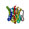
| ||||||||
|---|---|---|---|---|---|---|---|---|---|
| 1 |
| ||||||||
| Unit cell |
|
-Components
| #1: Protein | Transport protein Mass: 33311.801 Da / Num. of mol.: 1 / Mutation: Y113C, P190C Source method: isolated from a genetically manipulated source Source: (gene. exp.) Yersinia frederiksenii (bacteria) / Gene: NCTC11470_02445 / Production host: Escherichia coli (E. coli) / References: UniProt: A0A380PV03 | ||||
|---|---|---|---|---|---|
| #2: Chemical | ChemComp-CIT / Citric acid  Mass: 192.124 Da / Num. of mol.: 1 / Source method: obtained synthetically / Formula: C6H8O7 / Feature type: SUBJECT OF INVESTIGATION Mass: 192.124 Da / Num. of mol.: 1 / Source method: obtained synthetically / Formula: C6H8O7 / Feature type: SUBJECT OF INVESTIGATION | ||||
| #3: Chemical |   Mass: 356.540 Da / Num. of mol.: 2 / Source method: obtained synthetically / Formula: C21H40O4 Mass: 356.540 Da / Num. of mol.: 2 / Source method: obtained synthetically / Formula: C21H40O4#4: Water | ChemComp-HOH / | Water Mass: 18.015 Da / Num. of mol.: 16 / Source method: isolated from a natural source / Formula: H2O Mass: 18.015 Da / Num. of mol.: 16 / Source method: isolated from a natural source / Formula: H2OHas ligand of interest | Y | |
-Experimental details
-Experiment
| Experiment | Method: X-RAY DIFFRACTION / Number of used crystals: 1 |
|---|
- Sample preparation
| Crystal | Density Matthews: 2.42 Å3/Da / Density % sol: 49.17 % |
|---|---|
| Crystal grow | Temperature: 293.15 K / Method: lipidic cubic phase / pH: 5 Details: 0.2 M NH4H2PO4, 0.1 M (NH4)2SO4, 0.1 M Na+-citrate pH 5.0, 34% PEG 400, 4% 1,3-Butanediol |
-Data collection
| Diffraction | Mean temperature: 100 K / Serial crystal experiment: N | ||||||||||||||||||||||||||||||
|---|---|---|---|---|---|---|---|---|---|---|---|---|---|---|---|---|---|---|---|---|---|---|---|---|---|---|---|---|---|---|---|
| Diffraction source | Source: SYNCHROTRON / Site: SSRF / Beamline: BL19U1 / Wavelength: 0.97852 Å | ||||||||||||||||||||||||||||||
| Detector | Type: DECTRIS PILATUS3 6M / Detector: PIXEL / Date: Mar 23, 2019 | ||||||||||||||||||||||||||||||
| Radiation | Protocol: SINGLE WAVELENGTH / Monochromatic (M) / Laue (L): M / Scattering type: x-ray | ||||||||||||||||||||||||||||||
| Radiation wavelength | Wavelength: 0.97852 Å / Relative weight: 1 | ||||||||||||||||||||||||||||||
| Reflection | Resolution: 2.851→48.279 Å / Num. obs: 7621 / % possible obs: 96.1 % / Redundancy: 3.6 % / Biso Wilson estimate: 47.59 Å2 / CC1/2: 0.991 / Rmerge(I) obs: 0.145 / Rpim(I) all: 0.087 / Rrim(I) all: 0.171 / Net I/σ(I): 7.6 / Num. measured all: 27463 | ||||||||||||||||||||||||||||||
| Reflection shell | Diffraction-ID: 1
|
-Phasing
| Phasing | Method: molecular replacement |
|---|
- Processing
| Software |
| ||||||||||||||||||||||||
|---|---|---|---|---|---|---|---|---|---|---|---|---|---|---|---|---|---|---|---|---|---|---|---|---|---|
| Refinement | Method to determine structure: MOLECULAR REPLACEMENT Starting model: 4N7X Resolution: 2.861→48.279 Å / SU ML: 0.36 / Cross valid method: THROUGHOUT / σ(F): 1.34 / Phase error: 27.94 / Stereochemistry target values: ML
| ||||||||||||||||||||||||
| Solvent computation | Shrinkage radii: 0.9 Å / VDW probe radii: 1.11 Å / Solvent model: FLAT BULK SOLVENT MODEL | ||||||||||||||||||||||||
| Displacement parameters | Biso max: 89.3 Å2 / Biso mean: 42.6438 Å2 / Biso min: 19.08 Å2 | ||||||||||||||||||||||||
| Refinement step | Cycle: final / Resolution: 2.861→48.279 Å
| ||||||||||||||||||||||||
| LS refinement shell | Refine-ID: X-RAY DIFFRACTION / Rfactor Rfree error: 0
|
