 Movie
Movie Controller

[English] 日本語
 Yorodumi
- PDB-3k2g: Crystal structure of a Resiniferatoxin-binding protein from Rhodo... -
+ Open data

- Basic information
| Entry | Database: PDB / ID: 3k2g | ||||||
|---|---|---|---|---|---|---|---|
| Title | Crystal structure of a Resiniferatoxin-binding protein from Rhodobacter sphaeroides | ||||||
 Components Components | Resiniferatoxin-binding, phosphotriesterase-related protein | ||||||
 Keywords Keywords | Resiniferatoxin binding protein / Resiniferatoxin-binding /  phosphotriesterase / TIM BARREL / Binuclear Zinc / PROTEIN STRUCTURE INITIATIVE II (PSI II) / STRUCTURAL GENOMICS / NYSGXRC / 9588c / New York SGX Research Center for Structural Genomics phosphotriesterase / TIM BARREL / Binuclear Zinc / PROTEIN STRUCTURE INITIATIVE II (PSI II) / STRUCTURAL GENOMICS / NYSGXRC / 9588c / New York SGX Research Center for Structural Genomics | ||||||
| Function / homology |  Function and homology information Function and homology informationcatabolic process / phosphoric diester hydrolase activity / zinc ion bindingSimilarity search - Function | ||||||
| Biological species |  Rhodobacter sphaeroides 2.4.1 (bacteria) Rhodobacter sphaeroides 2.4.1 (bacteria) | ||||||
| Method | X-RAY DIFFRACTION / SYNCHROTRON / SAD / Resolution: 1.8 Å | ||||||
 Authors Authors | Kumaran, D. / Burley, S.K. / Swaminathan, S. / New York SGX Research Center for Structural Genomics (NYSGXRC) | ||||||
 Citation Citation | Journal: To be Published Title: Crystal structure of a Resiniferatoxin-binding protein from Rhodobacter sphaeroides Authors: Kumaran, D. / Burley, S.K. / Swaminathan, S. | ||||||
| History |
|
- Structure visualization
| Structure viewer | Molecule: MolmilJmol/JSmol |
|---|
- Downloads & links
-Download
| PDBx/mmCIF format | 3k2g.cif.gz | 296 KB | Display | PDBx/mmCIF format |
|---|---|---|---|---|
| PDB format | pdb3k2g.ent.gz | 251.2 KB | Display | PDB format |
| PDBx/mmJSON format | 3k2g.json.gz | Tree view | PDBx/mmJSON format | |
| Others |  Other downloads Other downloads |
-Validation report
| Arichive directory | https://data.pdbj.org/pub/pdb/validation_reports/k2/3k2gftp://data.pdbj.org/pub/pdb/validation_reports/k2/3k2g | HTTPS FTP |
|---|
-Related structure data
| Similar structure data | |
|---|---|
| Other databases |


-Links
 PDBj
PDBj

- Assembly
| Deposited unit | 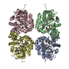
| ||||||||
|---|---|---|---|---|---|---|---|---|---|
| 1 |
| ||||||||
| 2 | 
| ||||||||
| 3 | 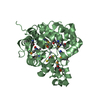
| ||||||||
| 4 | 
| ||||||||
| 5 | 
| ||||||||
| Unit cell |
| ||||||||
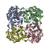
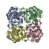
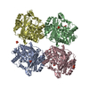
| Details | Authors state that the biological unit is tetramer. |
-Components
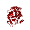
| #1: Protein | Mass: 40970.367 Da / Num. of mol.: 4 Source method: isolated from a genetically manipulated source Source: (gene. exp.) Rhodobacter sphaeroides 2.4.1 (bacteria)Strain: ATCC 17023 / 2.4.1 / NCIB 8253 / DSM 158 / Gene: RHOS4_37320, RHOS4_37320 (RSP_3690), RSP_3690 / Plasmid: BC-PSGX3 / Production host: Escherichia coli BL21(DE3) (bacteria) / Strain (production host): BL21(DE3) / References: UniProt: Q3IVY4#2: Chemical | ChemComp-ZN /   Mass: 65.409 Da / Num. of mol.: 8 / Source method: obtained synthetically / Formula: Zn Mass: 65.409 Da / Num. of mol.: 8 / Source method: obtained synthetically / Formula: Zn#3: Chemical | ChemComp-MG / |   Mass: 24.305 Da / Num. of mol.: 1 / Source method: obtained synthetically / Formula: Mg Mass: 24.305 Da / Num. of mol.: 1 / Source method: obtained synthetically / Formula: Mg#4: Chemical | ChemComp-DTV / ( Dithiothreitol  Mass: 154.251 Da / Num. of mol.: 4 / Source method: obtained synthetically / Formula: C4H10O2S2 Mass: 154.251 Da / Num. of mol.: 4 / Source method: obtained synthetically / Formula: C4H10O2S2#5: Water | ChemComp-HOH / | Water Mass: 18.015 Da / Num. of mol.: 872 / Source method: isolated from a natural source / Formula: H2O Mass: 18.015 Da / Num. of mol.: 872 / Source method: isolated from a natural source / Formula: H2O |
|---|
-Experimental details
-Experiment
| Experiment | Method: X-RAY DIFFRACTION / Number of used crystals: 1 |
|---|
- Sample preparation
| Crystal | Density Matthews: 2.33 Å3/Da / Density % sol: 47.24 % |
|---|---|
| Crystal grow | Temperature: 293 K / Method: vapor diffusion, sitting drop / pH: 8.5 Details: 25% PEG 3350, 0.2 M MgCl2, pH 8.5, VAPOR DIFFUSION, SITTING DROP, temperature 293K |
-Data collection
| Diffraction | Mean temperature: 100 K |
|---|---|
| Diffraction source | Source: SYNCHROTRON / Site: NSLS  / Beamline: X29A / Wavelength: 0.979 / Wavelength: 0.979 Å / Beamline: X29A / Wavelength: 0.979 / Wavelength: 0.979 Å |
| Detector | Type: ADSC QUANTUM 315 / Detector: CCD / Date: Sep 24, 2009 / Details: MIRRORS |
| Radiation | Monochromator: SI III / Protocol: SINGLE WAVELENGTH / Monochromatic (M) / Laue (L): M / Scattering type: x-ray |
| Radiation wavelength | Wavelength: 0.979 Å / Relative weight: 1 |
| Reflection | Resolution: 1.8→50 Å / Num. all: 127592 / Num. obs: 127592 / % possible obs: 90.7 % / Observed criterion σ(F): 0 / Observed criterion σ(I): 0 / Redundancy: 5.9 % / Rmerge(I) obs: 0.097 / Net I/σ(I): 17.8 |
| Reflection shell | Resolution: 1.8→1.86 Å / Redundancy: 2.3 % / Rmerge(I) obs: 0.3 / Mean I/σ(I) obs: 2 / Num. unique all: 7765 / % possible all: 55.7 |
- Processing
| Software |
| ||||||||||||||||||||||||||||||||||||||||||||||||||||||||||||
|---|---|---|---|---|---|---|---|---|---|---|---|---|---|---|---|---|---|---|---|---|---|---|---|---|---|---|---|---|---|---|---|---|---|---|---|---|---|---|---|---|---|---|---|---|---|---|---|---|---|---|---|---|---|---|---|---|---|---|---|---|---|
| Refinement | Method to determine structure: SAD / Resolution: 1.8→44.79 Å / Rfactor Rfree error: 0.003 / Data cutoff high absF: 121012.14 / Data cutoff low absF: 0 / Isotropic thermal model: RESTRAINED / Cross valid method: THROUGHOUT / σ(F): 0 / σ(I): 0 / Stereochemistry target values: Engh & Huber
| ||||||||||||||||||||||||||||||||||||||||||||||||||||||||||||
| Solvent computation | Solvent model: FLAT MODEL / Bsol: 40.783 Å2 / ksol: 0.360686 e/Å3 | ||||||||||||||||||||||||||||||||||||||||||||||||||||||||||||
| Displacement parameters | Biso mean: 22 Å2
| ||||||||||||||||||||||||||||||||||||||||||||||||||||||||||||
| Refine analyze |
| ||||||||||||||||||||||||||||||||||||||||||||||||||||||||||||
| Refinement step | Cycle: LAST / Resolution: 1.8→44.79 Å
| ||||||||||||||||||||||||||||||||||||||||||||||||||||||||||||
| Refine LS restraints |
| ||||||||||||||||||||||||||||||||||||||||||||||||||||||||||||
| Refine LS restraints NCS | NCS model details: NONE | ||||||||||||||||||||||||||||||||||||||||||||||||||||||||||||
| LS refinement shell | Resolution: 1.8→1.91 Å / Rfactor Rfree error: 0.012 / Total num. of bins used: 6
|
