 Movie
Movie Controller

[English] 日本語
 Yorodumi
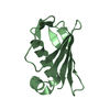
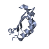
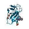
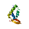
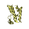
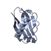
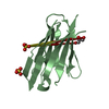
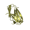
- PDB-6lg3: Crystal structure of a bacterial toxin from Mycobacterium tuberculosis -
+ Open data

- Basic information
| Entry | Database: PDB / ID: 6lg3 | ||||||
|---|---|---|---|---|---|---|---|
| Title | Crystal structure of a bacterial toxin from Mycobacterium tuberculosis | ||||||
 Components Components | PhiRv1 phage protein | ||||||
 Keywords Keywords |  TOXIN / bacterial toxin TOXIN / bacterial toxin | ||||||
| Function / homology | PhiRv1 phage protein / Probable PhiRv1 phage protein Function and homology information Function and homology information | ||||||
| Biological species |   Mycobacterium tuberculosis (bacteria) Mycobacterium tuberculosis (bacteria) | ||||||
| Method | X-RAY DIFFRACTION / SYNCHROTRON / MOLECULAR REPLACEMENT / Resolution: 1.90057258182 Å | ||||||
 Authors Authors | Zhou, J. / Jiang, Y. / Luo, Z. / Yin, L. | ||||||
| Funding support |  China, 1items China, 1items
| ||||||
 Citation Citation | Journal: Sci Adv / Year: 2020 Title: Mycobacterial EST12 activates a RACK1-NLRP3-gasdermin D pyroptosis-IL-1 beta immune pathway. Authors: Qu, Z. / Zhou, J. / Zhou, Y. / Xie, Y. / Jiang, Y. / Wu, J. / Luo, Z. / Liu, G. / Yin, L. / Zhang, X.L. | ||||||
| History |
|
- Structure visualization
| Structure viewer | Molecule: MolmilJmol/JSmol |
|---|
- Downloads & links
-Download
| PDBx/mmCIF format | 6lg3.cif.gz | 51.1 KB | Display | PDBx/mmCIF format |
|---|---|---|---|---|
| PDB format | pdb6lg3.ent.gz | 29 KB | Display | PDB format |
| PDBx/mmJSON format | 6lg3.json.gz | Tree view | PDBx/mmJSON format | |
| Others |  Other downloads Other downloads |
-Validation report
| Arichive directory | https://data.pdbj.org/pub/pdb/validation_reports/lg/6lg3ftp://data.pdbj.org/pub/pdb/validation_reports/lg/6lg3 | HTTPS FTP |
|---|
-Related structure data
| Related structure data |  2n9vS S: Starting model for refinement |
|---|---|
| Similar structure data |


-Links
 PDBj
PDBj- Assembly
| Deposited unit | 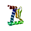
| ||||||||||||
|---|---|---|---|---|---|---|---|---|---|---|---|---|---|
| 1 |
| ||||||||||||
| Unit cell |
|
-Components
| #1: Protein | Mass: 8038.860 Da / Num. of mol.: 1 Source method: isolated from a genetically manipulated source Source: (gene. exp.) Mycobacterium tuberculosis (bacteria) / Production host: Escherichia coli (E. coli) / References: UniProt: A0A0T9XTX3, UniProt: O06611*PLUS |
|---|---|
| #2: Water | ChemComp-HOH / Water Mass: 18.015 Da / Num. of mol.: 37 / Source method: isolated from a natural source / Formula: H2O Mass: 18.015 Da / Num. of mol.: 37 / Source method: isolated from a natural source / Formula: H2O |
-Experimental details
-Experiment
| Experiment | Method: X-RAY DIFFRACTION / Number of used crystals: 1 |
|---|
- Sample preparation
| Crystal | Density Matthews: 2.66 Å3/Da / Density % sol: 53.71 % |
|---|---|
| Crystal grow | Temperature: 277 K / Method: vapor diffusion, sitting drop Details: 6% v/v Tacsimate pH 6.0, 0.1M MES monohydrate pH 6.0, 25% w/v Polyethylene glycol 4000 |
-Data collection
| Diffraction | Mean temperature: 95 K / Serial crystal experiment: N |
|---|---|
| Diffraction source | Source: SYNCHROTRON / Site: SSRF / Beamline: BL19U1 / Wavelength: 0.979 Å |
| Detector | Type: ADSC QUANTUM 315 / Detector: CCD / Date: May 10, 2016 |
| Radiation | Protocol: SINGLE WAVELENGTH / Monochromatic (M) / Laue (L): M / Scattering type: x-ray |
| Radiation wavelength | Wavelength: 0.979 Å / Relative weight: 1 |
| Reflection | Resolution: 1.9→19.35 Å / Num. obs: 12018 / % possible obs: 99 % / Redundancy: 1.9 % / Biso Wilson estimate: 33.1999350093 Å2 / Rmerge(I) obs: 0.143 / Net I/σ(I): 5.71 |
| Reflection shell | Resolution: 1.901→1.969 Å / Rmerge(I) obs: 0.213 / Mean I/σ(I) obs: 2.37 / Num. unique obs: 634 |
- Processing
| Software |
| ||||||||||||||||||||||||||||||||||||||||||||||||||||||||||||||||||||||
|---|---|---|---|---|---|---|---|---|---|---|---|---|---|---|---|---|---|---|---|---|---|---|---|---|---|---|---|---|---|---|---|---|---|---|---|---|---|---|---|---|---|---|---|---|---|---|---|---|---|---|---|---|---|---|---|---|---|---|---|---|---|---|---|---|---|---|---|---|---|---|---|
| Refinement | Method to determine structure: MOLECULAR REPLACEMENT Starting model: 2N9V Resolution: 1.90057258182→19.342176936 Å / SU ML: 0.207558956701 / Cross valid method: FREE R-VALUE / σ(F): 1.35089351612 / Phase error: 25.2504637423
| ||||||||||||||||||||||||||||||||||||||||||||||||||||||||||||||||||||||
| Solvent computation | Shrinkage radii: 0.9 Å / VDW probe radii: 1.11 Å | ||||||||||||||||||||||||||||||||||||||||||||||||||||||||||||||||||||||
| Displacement parameters | Biso mean: 39.7739305669 Å2 | ||||||||||||||||||||||||||||||||||||||||||||||||||||||||||||||||||||||
| Refinement step | Cycle: LAST / Resolution: 1.90057258182→19.342176936 Å
| ||||||||||||||||||||||||||||||||||||||||||||||||||||||||||||||||||||||
| Refine LS restraints |
| ||||||||||||||||||||||||||||||||||||||||||||||||||||||||||||||||||||||
| LS refinement shell |
| ||||||||||||||||||||||||||||||||||||||||||||||||||||||||||||||||||||||
| Refinement TLS params. | Method: refined / Origin x: 17.8878586915 Å / Origin y: 29.682154433 Å / Origin z: -0.393770085368 Å
| ||||||||||||||||||||||||||||||||||||||||||||||||||||||||||||||||||||||
| Refinement TLS group | Selection details: all |
