 Movie
Movie Controller

[English] 日本語
 Yorodumi
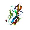
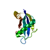
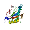
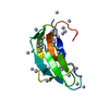
- PDB-4aix: Crystallographic structure of an amyloidogenic variant, 3rC34Y, o... -
+ Open data

- Basic information
| Entry | Database: PDB / ID: 4aix | ||||||
|---|---|---|---|---|---|---|---|
| Title | Crystallographic structure of an amyloidogenic variant, 3rC34Y, of the germinal line lambda 3 | ||||||
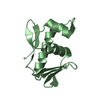
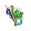
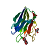
 Components Components | IG LAMBDA CHAIN V-IV REGION BAU | ||||||
 Keywords Keywords |  IMMUNE SYSTEM / AMYLOIDOSIS IMMUNE SYSTEM / AMYLOIDOSIS | ||||||
| Function / homology | Immunoglobulins / Immunoglobulin-like / Sandwich / Mainly Beta Function and homology information Function and homology information | ||||||
| Biological species |  HOMO SAPIENS (human) HOMO SAPIENS (human) | ||||||
| Method | X-RAY DIFFRACTION / SYNCHROTRON / MOLECULAR REPLACEMENT / Resolution: 1.8 Å | ||||||
 Authors Authors | Villalba, M.I. / Luna, O.D. / Rudino-Pinera, E. / Sanchez, R. / Sanchez-Lopez, R. / Rojas-Trejo, S. / Olamendi-Portugal, T. / Fernandez-Velasco, D.A. / Becerril, B. | ||||||
 Citation Citation | Journal: J.Biol.Chem. / Year: 2015 Title: Site-Directed Mutagenesis Reveals Regions Implicated in the Stability and Fiber Formation of Human Lambda3R Light Chains. Authors: Villalba, M.I. / Canul-Tec, J.C. / Luna-Martinez, O.D. / Sanchez-Alcala, R. / Olamendi-Portugal, T. / Rudino-Pinera, E. / Rojas, S. / Sanchez-Lopez, R. / Fernandez-Velasco, D.A. / Becerril, B. | ||||||
| History |
|
- Structure visualization
| Structure viewer | Molecule: MolmilJmol/JSmol |
|---|
- Downloads & links
-Download
| PDBx/mmCIF format | 4aix.cif.gz | 101.3 KB | Display | PDBx/mmCIF format |
|---|---|---|---|---|
| PDB format | pdb4aix.ent.gz | 79.1 KB | Display | PDB format |
| PDBx/mmJSON format | 4aix.json.gz | Tree view | PDBx/mmJSON format | |
| Others |  Other downloads Other downloads |
-Validation report
| Arichive directory | https://data.pdbj.org/pub/pdb/validation_reports/ai/4aixftp://data.pdbj.org/pub/pdb/validation_reports/ai/4aix | HTTPS FTP |
|---|
-Related structure data
| Related structure data |  4aizC 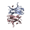 4aj0C  1lilS C: citing same article ( S: Starting model for refinement |
|---|---|
| Similar structure data |
-Links
 PDBj
PDBj

- Assembly
| Deposited unit | 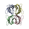
| ||||||||
|---|---|---|---|---|---|---|---|---|---|
| 1 | 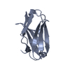
| ||||||||
| 2 | 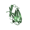
| ||||||||
| 3 | 
| ||||||||
| 4 | 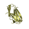
| ||||||||
| Unit cell |
|
-Components
| #1: Antibody | Mass: 11403.485 Da / Num. of mol.: 4 Source method: isolated from a genetically manipulated source Source: (gene. exp.) HOMO SAPIENS (human) / Production host:  ESCHERICHIA COLI (E. coli) / Strain (production host): BL21(DE3) ESCHERICHIA COLI (E. coli) / Strain (production host): BL21(DE3)#2: Water | ChemComp-HOH / | Water Mass: 18.015 Da / Num. of mol.: 356 / Source method: isolated from a natural source / Formula: H2O Mass: 18.015 Da / Num. of mol.: 356 / Source method: isolated from a natural source / Formula: H2OSequence details | BECAUSE THIS VARIANT IS RESULT OF MODIFICATIONS OVER 3RJL2 IT IS NOT DEPOSITED IN ANY DATA BASE. ...BECAUSE THIS VARIANT IS RESULT OF MODIFICATI | |
|---|
-Experimental details
-Experiment
| Experiment | Method: X-RAY DIFFRACTION / Number of used crystals: 1 |
|---|
- Sample preparation
| Crystal | Density Matthews: 1.86 Å3/Da / Density % sol: 33.96 % / Description: NONE |
|---|---|
| Crystal grow | pH: 3 / Details: 2.3 M AMMONIUM SULFATE, 40 % TREHALOSE |
-Data collection
| Diffraction | Mean temperature: 100 K |
|---|---|
| Diffraction source | Source: SYNCHROTRON / Site: NSLS  / Beamline: X6A / Wavelength: 0.972 / Beamline: X6A / Wavelength: 0.972 |
| Detector | Type: ADSC QUANTUM 210 / Detector: CCD / Date: May 1, 2011 Details: DOUBLE CRYSTAL CHANNEL CUT, SI(111), 1M LONG RH COATED TOROIDAL MIRROR FOR VERTICAL AND HORIZONTAL FOCUSING |
| Radiation | Protocol: SINGLE WAVELENGTH / Monochromatic (M) / Laue (L): M / Scattering type: x-ray |
| Radiation wavelength | Wavelength: 0.972 Å / Relative weight: 1 |
| Reflection | Resolution: 1.8→27.65 Å / Num. obs: 31131 / % possible obs: 100 % / Observed criterion σ(I): 0 / Redundancy: 5.7 % / Biso Wilson estimate: 16.27 Å2 / Rmerge(I) obs: 0.06 / Net I/σ(I): 10.4 |
| Reflection shell | Resolution: 1.8→1.9 Å / Redundancy: 5.7 % / Rmerge(I) obs: 0.32 / Mean I/σ(I) obs: 2.3 / % possible all: 100 |
- Processing
| Software |
| ||||||||||||||||||||||||||||||||||||||||||||||||||||||||||||||||||||||||||||||||||||
|---|---|---|---|---|---|---|---|---|---|---|---|---|---|---|---|---|---|---|---|---|---|---|---|---|---|---|---|---|---|---|---|---|---|---|---|---|---|---|---|---|---|---|---|---|---|---|---|---|---|---|---|---|---|---|---|---|---|---|---|---|---|---|---|---|---|---|---|---|---|---|---|---|---|---|---|---|---|---|---|---|---|---|---|---|---|
| Refinement | Method to determine structure: MOLECULAR REPLACEMENT Starting model: PDB ENTRY 1LIL Resolution: 1.8→24.869 Å / SU ML: 0.48 / σ(F): 2.85 / Phase error: 20.93 / Stereochemistry target values: ML
| ||||||||||||||||||||||||||||||||||||||||||||||||||||||||||||||||||||||||||||||||||||
| Solvent computation | Shrinkage radii: 0.61 Å / VDW probe radii: 0.9 Å / Solvent model: FLAT BULK SOLVENT MODEL / Bsol: 45.558 Å2 / ksol: 0.398 e/Å3 | ||||||||||||||||||||||||||||||||||||||||||||||||||||||||||||||||||||||||||||||||||||
| Displacement parameters | Biso mean: 20.87 Å2
| ||||||||||||||||||||||||||||||||||||||||||||||||||||||||||||||||||||||||||||||||||||
| Refinement step | Cycle: LAST / Resolution: 1.8→24.869 Å
| ||||||||||||||||||||||||||||||||||||||||||||||||||||||||||||||||||||||||||||||||||||
| Refine LS restraints |
| ||||||||||||||||||||||||||||||||||||||||||||||||||||||||||||||||||||||||||||||||||||
| LS refinement shell |
|
