 Movie
Movie Controller

[English] 日本語
 Yorodumi
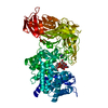
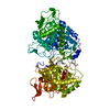
- PDB-6j63: Crystal structure of Arabidopsis thaliana HPPD complexed with NTBC -
+ Open data

- Basic information
| Entry | Database: PDB / ID: 6j63 | |||||||||
|---|---|---|---|---|---|---|---|---|---|---|
| Title | Crystal structure of Arabidopsis thaliana HPPD complexed with NTBC | |||||||||
 Components Components | 4-hydroxyphenylpyruvate dioxygenase | |||||||||
 Keywords Keywords | OXIDOREDUCTASE / 4-hydroxyphenylpyruvate dioxygenase / nitisinone / type I tyrosinemia / drug discovery | |||||||||
| Function / homology |  Function and homology information4-hydroxyphenylpyruvate dioxygenase / 4-hydroxyphenylpyruvate dioxygenase activity / tyrosine catabolic process / L-phenylalanine catabolic process / iron ion binding / identical protein binding / cytoplasm Function and homology information4-hydroxyphenylpyruvate dioxygenase / 4-hydroxyphenylpyruvate dioxygenase activity / tyrosine catabolic process / L-phenylalanine catabolic process / iron ion binding / identical protein binding / cytoplasmSimilarity search - Function | |||||||||
| Biological species |  Arabidopsis thaliana (thale cress) Arabidopsis thaliana (thale cress) | |||||||||
| Method | X-RAY DIFFRACTION / SYNCHROTRON / MOLECULAR REPLACEMENT / Resolution: 2.624 Å | |||||||||
 Authors Authors | Yang, W.C. / Yang, G.F. | |||||||||
| Funding support |  China, 1items China, 1items
| |||||||||
 Citation Citation | Journal: FEBS J. / Year: 2019 Title: Molecular insights into the mechanism of 4-hydroxyphenylpyruvate dioxygenase inhibition: enzyme kinetics, X-ray crystallography and computational simulations. Authors: Lin, H.Y. / Yang, J.F. / Wang, D.W. / Hao, G.F. / Dong, J.Q. / Wang, Y.X. / Yang, W.C. / Wu, J.W. / Zhan, C.G. / Yang, G.F. | |||||||||
| History |
|
- Structure visualization
| Structure viewer | Molecule: MolmilJmol/JSmol |
|---|
- Downloads & links
-Download
| PDBx/mmCIF format | 6j63.cif.gz | 285.4 KB | Display | PDBx/mmCIF format |
|---|---|---|---|---|
| PDB format | pdb6j63.ent.gz | 226.1 KB | Display | PDB format |
| PDBx/mmJSON format | 6j63.json.gz | Tree view | PDBx/mmJSON format | |
| Others |  Other downloads Other downloads |
-Validation report
| Arichive directory | https://data.pdbj.org/pub/pdb/validation_reports/j6/6j63ftp://data.pdbj.org/pub/pdb/validation_reports/j6/6j63 | HTTPS FTP |
|---|
-Related structure data
| Related structure data |  5ywgC 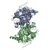 6isdC 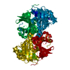 1sqdS S: Starting model for refinement C: citing same article ( |
|---|---|
| Similar structure data |


-Links
 PDBj
PDBj- Assembly
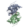
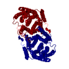
| Deposited unit | 
| ||||||||
|---|---|---|---|---|---|---|---|---|---|
| 1 | 
| ||||||||
| 2 | 
| ||||||||
| Unit cell |
|
-Components
| #1: Protein | / 4-hydroxyphenylpyruvic acid oxidase / HPPDase Mass: 48873.832 Da / Num. of mol.: 4 Source method: isolated from a genetically manipulated source Source: (gene. exp.) Arabidopsis thaliana (thale cress) / Gene: HPDProduction host:  Escherichia coli 'BL21-Gold(DE3)pLysS AG' (bacteria) Escherichia coli 'BL21-Gold(DE3)pLysS AG' (bacteria)Strain (production host): BL21-Gold(DE3)pLysS AG References: UniProt: P93836, 4-hydroxyphenylpyruvate dioxygenase#2: Chemical | ChemComp-FE / Iron  Mass: 55.845 Da / Num. of mol.: 4 / Source method: obtained synthetically / Formula: Fe Mass: 55.845 Da / Num. of mol.: 4 / Source method: obtained synthetically / Formula: Fe#3: Chemical | ChemComp-NTD /   Mass: 329.228 Da / Num. of mol.: 4 / Source method: obtained synthetically / Formula: C14H10F3NO5 Mass: 329.228 Da / Num. of mol.: 4 / Source method: obtained synthetically / Formula: C14H10F3NO5#4: Water | ChemComp-HOH / | Water Mass: 18.015 Da / Num. of mol.: 18 / Source method: isolated from a natural source / Formula: H2O Mass: 18.015 Da / Num. of mol.: 18 / Source method: isolated from a natural source / Formula: H2O |
|---|
-Experimental details
-Experiment
| Experiment | Method: X-RAY DIFFRACTION / Number of used crystals: 1 |
|---|
- Sample preparation
| Crystal | Density Matthews: 2.3 Å3/Da / Density % sol: 46.48 % |
|---|---|
| Crystal grow | Temperature: 291.16 K / Method: vapor diffusion, hanging drop / pH: 4.5 / Details: 0.1M NaAc at pH 4.5, 0.1M NaCl, 40% PEG 400 |
-Data collection
| Diffraction | Mean temperature: 100 K / Serial crystal experiment: N |
|---|---|
| Diffraction source | Source: SYNCHROTRON / Site: SSRF / Beamline: BL17U1 / Wavelength: 0.9796 Å |
| Detector | Type: ADSC QUANTUM 315r / Detector: CCD / Date: May 23, 2015 |
| Radiation | Protocol: SINGLE WAVELENGTH / Monochromatic (M) / Laue (L): M / Scattering type: x-ray |
| Radiation wavelength | Wavelength: 0.9796 Å / Relative weight: 1 |
| Reflection | Resolution: 2.62→50 Å / Num. obs: 52627 / % possible obs: 99.1 % / Redundancy: 3.7 % / CC1/2: 0.764 / Rmerge(I) obs: 0.571 / Rsym value: 0.571 / Net I/σ(I): 9.72 |
| Reflection shell | Resolution: 2.62→2.77 Å / Redundancy: 3.6 % / Rmerge(I) obs: 1.687 / Num. unique obs: 7642 / CC1/2: 0.429 / Rsym value: 1.687 / % possible all: 98.9 |
- Processing
| Software |
| ||||||||||||||||||||||||||||||||||||||||||||||||||||||||||||||||||||||||||||||||||||||||||||||||||||||||||||||||||||||||||||||||||||||||||||
|---|---|---|---|---|---|---|---|---|---|---|---|---|---|---|---|---|---|---|---|---|---|---|---|---|---|---|---|---|---|---|---|---|---|---|---|---|---|---|---|---|---|---|---|---|---|---|---|---|---|---|---|---|---|---|---|---|---|---|---|---|---|---|---|---|---|---|---|---|---|---|---|---|---|---|---|---|---|---|---|---|---|---|---|---|---|---|---|---|---|---|---|---|---|---|---|---|---|---|---|---|---|---|---|---|---|---|---|---|---|---|---|---|---|---|---|---|---|---|---|---|---|---|---|---|---|---|---|---|---|---|---|---|---|---|---|---|---|---|---|---|---|
| Refinement | Method to determine structure: MOLECULAR REPLACEMENT Starting model: 1SQD Resolution: 2.624→29.187 Å / SU ML: 0.51 / Cross valid method: FREE R-VALUE / σ(F): 1.35 / Phase error: 31.44 / Stereochemistry target values: ML
| ||||||||||||||||||||||||||||||||||||||||||||||||||||||||||||||||||||||||||||||||||||||||||||||||||||||||||||||||||||||||||||||||||||||||||||
| Solvent computation | Shrinkage radii: 0.9 Å / VDW probe radii: 1.11 Å / Solvent model: FLAT BULK SOLVENT MODEL | ||||||||||||||||||||||||||||||||||||||||||||||||||||||||||||||||||||||||||||||||||||||||||||||||||||||||||||||||||||||||||||||||||||||||||||
| Refinement step | Cycle: LAST / Resolution: 2.624→29.187 Å
| ||||||||||||||||||||||||||||||||||||||||||||||||||||||||||||||||||||||||||||||||||||||||||||||||||||||||||||||||||||||||||||||||||||||||||||
| Refine LS restraints |
| ||||||||||||||||||||||||||||||||||||||||||||||||||||||||||||||||||||||||||||||||||||||||||||||||||||||||||||||||||||||||||||||||||||||||||||
| LS refinement shell |
|
