 Movie
Movie Controller


+ Open data

- Basic information
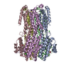
| Entry | Database: PDB / ID: 6ivl | ||||||
|---|---|---|---|---|---|---|---|
| Title | Crystal structure of a membrane protein L259A | ||||||
 Components Components | Ibestrophin | ||||||
 Keywords Keywords |  MEMBRANE PROTEIN MEMBRANE PROTEIN | ||||||
| Function / homology | UPF0187 family / Bestrophin/UPF0187 / Bestrophin, RFP-TM, chloride channel / chloride channel activity / membrane / plasma membrane / ACETIC ACID / Ibestrophin / Ibestrophin Function and homology information Function and homology information | ||||||
| Biological species |  Klebsiella pneumoniae (bacteria) Klebsiella pneumoniae (bacteria) | ||||||
| Method | X-RAY DIFFRACTION / SYNCHROTRON / MOLECULAR REPLACEMENT / Resolution: 3.4 Å | ||||||
 Authors Authors | Kittredge, A. / Fukuda, F. / Zhang, Y. / Yang, T. | ||||||
| Funding support |  United States, 1items United States, 1items
| ||||||
 Citation Citation | Journal: Commun Biol / Year: 2019 Title: Dual Ca2+-dependent gates in human Bestrophin1 underlie disease-causing mechanisms of gain-of-function mutations. Authors: Ji, C. / Kittredge, A. / Hopiavuori, A. / Ward, N. / Chen, S. / Fukuda, Y. / Zhang, Y. / Yang, T. | ||||||
| History |
|
- Structure visualization
| Structure viewer | Molecule: MolmilJmol/JSmol |
|---|
- Downloads & links
-Download
| PDBx/mmCIF format | 6ivl.cif.gz | 274.5 KB | Display | PDBx/mmCIF format |
|---|---|---|---|---|
| PDB format | pdb6ivl.ent.gz | 222.1 KB | Display | PDB format |
| PDBx/mmJSON format | 6ivl.json.gz | Tree view | PDBx/mmJSON format | |
| Others |  Other downloads Other downloads |
-Validation report
| Arichive directory | https://data.pdbj.org/pub/pdb/validation_reports/iv/6ivlftp://data.pdbj.org/pub/pdb/validation_reports/iv/6ivl | HTTPS FTP |
|---|
-Related structure data
| Related structure data |  6iv0C 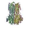 6iv1C 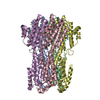 6iv2C 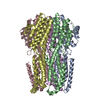 6iv3C  6iv4C  6ivjC  6ivkC 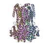 6ivmC 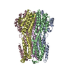 6ivnC 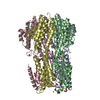 6ivoC  6ivpC 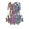 6ivqC  6ivrC  6ivwC  6jlfC C: citing same article ( |
|---|---|
| Similar structure data |


-Links
 PDBj
PDBj

- Assembly
| Deposited unit | 
| ||||||||
|---|---|---|---|---|---|---|---|---|---|
| 1 |
| ||||||||
| Unit cell |
|
-Components
| #1: Protein | Mass: 33789.164 Da / Num. of mol.: 5 / Mutation: L259A Source method: isolated from a genetically manipulated source Source: (gene. exp.) Klebsiella pneumoniae (bacteria)Gene: yneE, AGG09_26735, B1727_16705, B4U21_14105, B4U25_16730, B4U30_16350, B4U35_20780, BN49_2925, C3483_12335, C7V41_19985, CPT10_17935, CWN54_25435, D0897_06110, DXF97_13395, DY552_11160, DZB15_ ...Gene: yneE, AGG09_26735, B1727_16705, B4U21_14105, B4U25_16730, B4U30_16350, B4U35_20780, BN49_2925, C3483_12335, C7V41_19985, CPT10_17935, CWN54_25435, D0897_06110, DXF97_13395, DY552_11160, DZB15_11820, NCTC11679_02573, NCTC13465_00112, NCTC5052_01714, NCTC8849_03195, NCTC9637_03467, NCTC9645_05950, NCTC9661_03571, SAMEA104305404_11875, SAMEA23986918_00256, SAMEA24002668_02597, SAMEA3649709_04169, SAMEA4394730_00268 Production host: Escherichia coli (E. coli) / References: UniProt: W9BH30, UniProt: W1ELP7*PLUS#2: Chemical | ChemComp-ZN /   Mass: 65.409 Da / Num. of mol.: 16 / Source method: obtained synthetically / Formula: Zn Mass: 65.409 Da / Num. of mol.: 16 / Source method: obtained synthetically / Formula: Zn#3: Chemical | ChemComp-CL / Chloride  Mass: 35.453 Da / Num. of mol.: 6 / Source method: obtained synthetically / Formula: Cl Mass: 35.453 Da / Num. of mol.: 6 / Source method: obtained synthetically / Formula: Cl#4: Chemical | ChemComp-ACY / | Acetic acid  Mass: 60.052 Da / Num. of mol.: 1 / Source method: obtained synthetically / Formula: C2H4O2 Mass: 60.052 Da / Num. of mol.: 1 / Source method: obtained synthetically / Formula: C2H4O2#5: Water | ChemComp-HOH / | Water Mass: 18.015 Da / Num. of mol.: 10 / Source method: isolated from a natural source / Formula: H2O Mass: 18.015 Da / Num. of mol.: 10 / Source method: isolated from a natural source / Formula: H2O |
|---|
-Experimental details
-Experiment
| Experiment | Method: X-RAY DIFFRACTION / Number of used crystals: 1 |
|---|
- Sample preparation
| Crystal | Density Matthews: 4.4 Å3/Da / Density % sol: 72.03 % |
|---|---|
| Crystal grow | Temperature: 293 K / Method: vapor diffusion / pH: 6 Details: 0.05 M zinc acetate, 6% v/v ethylene glycol, 0.1 M sodium cacodylate, pH 6.0, 6.6 % w/v PEG 8000 |
-Data collection
| Diffraction | Mean temperature: 100 K / Serial crystal experiment: N |
|---|---|
| Diffraction source | Source: SYNCHROTRON / Site: APS / Beamline: 24-ID-C / Wavelength: 0.9791 Å |
| Detector | Type: ADSC QUANTUM 315 / Detector: CCD / Date: Nov 1, 2017 |
| Radiation | Protocol: SINGLE WAVELENGTH / Monochromatic (M) / Laue (L): M / Scattering type: x-ray |
| Radiation wavelength | Wavelength: 0.9791 Å / Relative weight: 1 |
| Reflection | Resolution: 3.4→161.9 Å / Num. obs: 41709 / % possible obs: 100 % / Redundancy: 6.6 % / Rmerge(I) obs: 0.088 / Rpim(I) all: 0.037 / Net I/σ(I): 15.4 |
| Reflection shell | Resolution: 3.4→3.54 Å / Redundancy: 6.8 % / Rmerge(I) obs: 0.835 / Mean I/σ(I) obs: 2.6 / Num. unique obs: 4641 / CC1/2: 0.757 / Rpim(I) all: 0.345 / % possible all: 100 |
- Processing
| Software |
| ||||||||||||||||||||||||||||||||||||||||||||||||||||||||||||||||||||||||||||||||||||||||||||||||||||||||||||||||||||||||||||||||||||||||||||||||||||||||||||||||||||||||||||||||||||||
|---|---|---|---|---|---|---|---|---|---|---|---|---|---|---|---|---|---|---|---|---|---|---|---|---|---|---|---|---|---|---|---|---|---|---|---|---|---|---|---|---|---|---|---|---|---|---|---|---|---|---|---|---|---|---|---|---|---|---|---|---|---|---|---|---|---|---|---|---|---|---|---|---|---|---|---|---|---|---|---|---|---|---|---|---|---|---|---|---|---|---|---|---|---|---|---|---|---|---|---|---|---|---|---|---|---|---|---|---|---|---|---|---|---|---|---|---|---|---|---|---|---|---|---|---|---|---|---|---|---|---|---|---|---|---|---|---|---|---|---|---|---|---|---|---|---|---|---|---|---|---|---|---|---|---|---|---|---|---|---|---|---|---|---|---|---|---|---|---|---|---|---|---|---|---|---|---|---|---|---|---|---|---|---|
| Refinement | Method to determine structure: MOLECULAR REPLACEMENT / Resolution: 3.4→48.8 Å / Cor.coef. Fo:Fc: 0.892 / Cor.coef. Fo:Fc free: 0.854 / Cross valid method: THROUGHOUT / ESU R Free: 0.439 / Details: HYDROGENS HAVE BEEN USED IF PRESENT IN THE INPUT
| ||||||||||||||||||||||||||||||||||||||||||||||||||||||||||||||||||||||||||||||||||||||||||||||||||||||||||||||||||||||||||||||||||||||||||||||||||||||||||||||||||||||||||||||||||||||
| Solvent computation | Ion probe radii: 0.8 Å / Shrinkage radii: 0.8 Å / VDW probe radii: 1.2 Å | ||||||||||||||||||||||||||||||||||||||||||||||||||||||||||||||||||||||||||||||||||||||||||||||||||||||||||||||||||||||||||||||||||||||||||||||||||||||||||||||||||||||||||||||||||||||
| Displacement parameters | Biso mean: 103.86 Å2
| ||||||||||||||||||||||||||||||||||||||||||||||||||||||||||||||||||||||||||||||||||||||||||||||||||||||||||||||||||||||||||||||||||||||||||||||||||||||||||||||||||||||||||||||||||||||
| Refinement step | Cycle: 1 / Resolution: 3.4→48.8 Å
| ||||||||||||||||||||||||||||||||||||||||||||||||||||||||||||||||||||||||||||||||||||||||||||||||||||||||||||||||||||||||||||||||||||||||||||||||||||||||||||||||||||||||||||||||||||||
| Refine LS restraints |
|